Korean J Urol.
2007 Feb;48(2):176-182. 10.4111/kju.2007.48.2.176.
Predicting Factors for Recurrence in First-time Stone Formers
- Affiliations
-
- 1Department of Urology, Chungbuk National University College of Medicine, Cheongju, Korea. lscuro@chungbuk.ac.kr
- KMID: 2139757
- DOI: http://doi.org/10.4111/kju.2007.48.2.176
Abstract
-
PURPOSE: The purpose of the present study was to determine the risk factors for predicting the recurrence in first-time urinary stone formers.
MATERIALS AND METHODS
A total of 121 patients, who presented at our hospital with first-time urinary stone episodes, between 1996 and 2005, and followed up for at least 3 years, were retrospectively evaluated. Of these, 65 patients (41 males, 24 females) recurred (R group) and 56 (38 males, 18 females) not (NR group) during the follow-up period. The blood chemistry and urinary analytes values, as well as the clinical characteristics between the NR and R groups were compared by gender. p-values less than 0.05 were used to indicate statistical significance.
RESULTS
There were no differences in the clinical characteristics between the NR and R groups. A comparison of the blood chemistry showed differences in the phosphate and calcium in men and women (p=0.047 and 0.034), respectively. Greater urinary excretion of phosphates were found in the R group than in the NR group (p=0.018), but was more prominent in men (p=0.006). No significant differences were found between the two groups with regard to metabolic abnormalities. A multivariate analysis revealed that urinary phosphates excretion was the sole predictor for stone recurrence (Exp beta=8.347, p=0.033). CONCLISIONS: Our results suggested that the increased excretion of urinary phosphates was a significant risk factor for stone recurrence, which might be useful as a prognostic marker.
Keyword
MeSH Terms
Figure
-
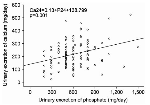
Fig. 1 Scatter plot shows a positive correlation between the 24-hour urinary excretions of phosphorus and calcium in first urinary stone formers.
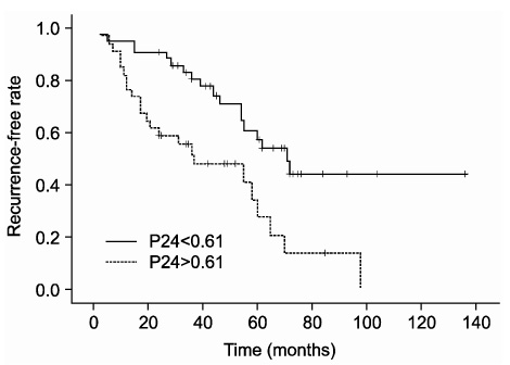
Fig. 2 Recurrence-free rate after first urinary stone, as calculated using the Kaplan-Meier curve method, in relation to urinary phosphate excretion (cut-off value=0.61, p=0.001).
Reference
-
1. Leusmann DB, Blaschke R, Schmandt W. Results of 5,035 stone analysis: a contribution to epidemiology of urinary stone disease. Scand J Urol Nephrol. 1990. 24:205–210.2. Tomson CR. Prevention of recurrent calcium stones: a rational approach. Br J Urol. 1995. 76:419–424.3. Trinchieri A, Ostini F, Nespoli R, Rovera F, Montanari E, Zanetti G. A prospective study of recurrence rate and risk factors for recurrence after a first renal stone. J Urol. 1999. 162:27–30.4. Siener R. Impact dietary habits on stone incidence. Urol Res. 2006. 34:131–133.5. Laube N, Pullmann M. The use of risk indices: do they predic recurrence? Yes, they (at least some) do. Urol Res. 2006. 34:118–121.6. Yagisawa T, Chandohoke PS, Fan J. Metabolic risk factors in patients with first-time and recurrent stone formations as determined by comprehensive metabolic evaluation. Urology. 1998. 52:750–755.7. Oh SY, Moon YT. Comparison of stone metabolic risk factors in patients with first-time and recurrent stone formations. Korean J Urol. 2004. 45:551–556.8. Kwon OJ, Ahn SH. Comparison of the lithogenic risk factors for first time and recurrent stone-formers. Korean J Urol. 2006. 47:1093–1098.9. Borghi L, Meschi T, Amato F, Briganti A, Novarini A, Giannini A. Urinary volume, water and recurrences in idio pathic calcium nephrolithiasis: 5-year randomized prospective study. J Urol. 1996. 155:839–843.10. Lee SY, Lee SC, Kim WJ. Metabolic abnormalities of 24-hour urinary lithogenic factors in recurrent stone formers. Korean J Urol. 2001. 42:69–74.11. Robertson WG. Is prevention of stone recurrence financially worthwhile? Urol Res. 2006. 34:157–161.12. Laube N, Hergarten S, Hoppe B, Schmidt M, Hesse A. Determination of the calcium oxalate crystallization risk from urine samples-the BONN-Risk-Index in comparison to other risk formulas. J Urol. 2004. 172:355–359.13. Werness PG, Brown CM, Smith LH, Finlayson B. EQUIL 2: a basic computer program for the calculation of urinary saturation. J Urol. 1885. 134:1242–1244.14. Siener R, Glatz S, Nicolay C, Hesse A. Prospective study on the efficacy of a selective treatment and risk factors for relapse in recurrent calcium oxalate stone patients. Eur Urol. 2003. 44:467–474.15. Ala-Opas M, Lehtonen T. Recurrences of urolithiasis. Ann Chir Gynaecol. 1990. 79:50–53.16. Ljunghall S, Danielson BG. A prospective study of renal stone recurrences. Br J Urol. 1984. 56:122–124.17. Pak CY. Resnick MI, Pak CY, editors. Hypercalciuric calcium nephrolithiasis. Urolithiasis. 1990. Philadelphia: Saunders;35–63.18. Williams CP, Child DF, Hudson PR, Soysa LD, Davies GK, Davies MG, et al. Inappropriate phosphate excretion in idiopathic hypercalciuria: the key to a common cause and future treatment? J Clin Pathol. 1996. 49:881–888.19. Bushinsky DA, Asplin JR. Thiazides reduce brushite, but not calcium oxalate, supersaturation, and stone formation in genetic hypercalciuric stone-forming rats. J Am Soc Nephrol. 2005. 16:417–424.20. Aihara K, Byer KJ, Khan SR. Calcium phosphate-induced renal epithelial injury and stone formation: involvement of reactive oxygen species. Kidney Int. 2003. 64:1283–1291.21. Prie D, Ravery V, Boccon-Gibod L, Friedlander G. Frequency of renal phosphate leak among patients with calcium nephrolithiasis. Kidney Int. 2001. 60:272–276.22. Broadus AE, Insogna KL, Lang R, Mallette LE, Oren DA, Gertner JM, et al. A consideration of the hormonal basis and phosphate leak hypothesis of absorptive hypercalciuria. J Clin Endocrinol Metab. 1984. 58:161–169.23. Dagnone AJ, Norman RW. Serum phosphate in stone formers: What does a low serum phosphate level mean? J Urol. 2000. 163:21–23.
- Full Text Links
-
- Actions
-
Cited
- CITED
-
- Close
- Share
-
- Similar articles
-
- Comparison of the Lithogenic Risk Factors for First Time and Recurrent Stone-formers
- Study on Uric Acid Level in Urinary Calcium Stone Formers
- Study on Serum and Urinary Calcium Level and Serum Parathyroid Hormone in Patients with Urinary Stone
- Study on Serum and Urinary Uric Acid Level in Patients with Urinary Stone
- Comparison of Metabolic Risk Factors in Urolithiasis Patients according to Family History
