Retroperitoneal Synovial Sarcoma Manifested by Obstructive Jaundice in an Elderly Woman: Case Report
- Affiliations
-
- 1Department of Gastroenterology, Kyung Hee University Hospital at Gangdong, Kyung Hee University School of Medicine, Seoul, Korea. krjoo@khu.ac.kr
- 2Department of Pathology, Kyung Hee University Hospital at Gangdong, Kyung Hee University School of Medicine, Seoul, Korea.
- 3Department of Radiology, Kyung Hee University Hospital at Gangdong, Kyung Hee University School of Medicine, Seoul, Korea.
Abstract
- Synovial sarcoma is a rare type of soft tissue sarcoma that arises in tissues containing synovial fluid, usually in the extremities. It has only rare occurrence in the retroperitoneal space. Early detection of retroperitoneal synovial sarcoma is difficult, since the retroperitoneal space is highly expandable and deeply hidden. Furthermore, the presenting symptoms are often vague and nonspecific, and are related to the pressure on adjacent structures. In this study, we present an unusual case of retroperitoneal synovial sarcoma with obstructive jaundice due to intrabiliary blood clots caused by invasion of bile duct by tumor. The obstructive jaundice was relieved through endoscopic removal of the blood clots and insertion of a biliary stent.
Keyword
MeSH Terms
Figure
-

Fig. 1 (A) Axial image of abdomen computed tomography scan revealing the presence of a huge retroperitoneal mass (14×8.5 cm) with direct liver invasion (arrowheads) and compressed inferior vena cava (arrow). (B) Coronal image also reveals encircling of the extrahepatic bile duct by the tumor (arrows).

Fig. 2 Magnetic resonance cholangiopancreatography shows the presence of a filling defect (arrow) at the hilar and common hepatic duct level of the bile duct.

Fig. 3 Endoscopic retrograde cholangiopancreatography shows a tubular filling defect (arrow); we could verify that the filling defect was movable during the intrabiliary injection of the contrast media. Some fresh blood was discharged from the papillary orifice when contrast media was injected into the bile duct (arrow in the inset).
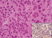
Fig. 4 Microscopic finding of the biopsied specimen reveals the presence of a tumor composed of infiltrating large epithelioid cells with marked nuclear pleomorphism and abundant eosinophilic cytoplasm (H&E stain, ×50). Immunostaining for CD99 shows positive reaction (inset).
Reference
-
1. Spillane AJ, A'Hern R, Judson IR, Fisher C, Thomas JM. Synovial sarcoma: a clinicopathologic, staging, and prognostic assessment. J Clin Oncol. 2000; 18:3794–3803. PMID: 11078492.
Article2. Pappo AS, Fontanesi J, Luo X, et al. Synovial sarcoma in children and adolescents: the St Jude Children's Research Hospital experience. J Clin Oncol. 1994; 12:2360–2366. PMID: 7964951.
Article3. Fisher C, Folpe AL, Hashimoto H, Weiss SW. Intra-abdominal synovial sarcoma: a clinicopathological study. Histopathology. 2004; 45:245–253. PMID: 15330802.
Article4. Pack GT, Tabah EJ. Primary retroperitoneal tumors: a study of 120 cases. Int Abstr Surg. 1954; 99:313–341. PMID: 13205424.5. Miyashita T, Imamura T, Ishikawa Y, Okinaga K, Kunii O, Miyashita H. Primary retroperitoneal synovial sarcoma. Intern Med. 1994; 33:692–696. PMID: 7849384.
Article6. Alhazzani AR, El-Sharkawy MS, Hassan H. Primary retroperitoneal synovial sarcoma in CT and MRI. Urol Ann. 2010; 2:39–41. PMID: 20842258.
Article
- Full Text Links
-
- Actions
-
Cited
- CITED
-
- Close
- Share
-
- Similar articles
-
- Primary Retroperitoneal Synovial Sarcoma: A Case Report
- Clinical Pathology, Catholic University Medical CollegeSynovial Sarcoma Manifested as a Subcutaneous Nodule of Lip: A case report
- Congenital Synovial Sarcoma in Ankle: Report of A Case
- A Case Report of Synovial Sarcoma
- Retroperitoneal Synovial Sarcoma: A case report
