Clin Endosc.
2014 Nov;47(6):568-570. 10.5946/ce.2014.47.6.568.
Endoscopic Diagnosis of Duodenal Stenosis in a 5-Month-Old Male Infant
- Affiliations
-
- 1Department of Pediatrics, Vanderbilt University School of Medicine, Nashville, TN, USA. michael.rosen@cchmc.org
- 2Department of Pediatric Surgery, Vanderbilt University School of Medicine, Nashville, TN, USA.
- 3Division of Gastroenterology, Hepatology and Nutrition, Cincinnati Children's Hospital Medical Center, Cincinnati, OH, USA.
- KMID: 2134497
- DOI: http://doi.org/10.5946/ce.2014.47.6.568
Abstract
- Duodenal stenosis and duodenal atresia are well-known gastrointestinal anomalies in patients with Down syndrome. Although duodenal atresia presents early and classically with vomiting in the immediate neonatal period, the presentation of duodenal stenosis can be significantly more subtle and the diagnosis delayed. Here, we describe the case of a 5-month-old male infant with Down syndrome and delayed presentation of high-grade duodenal stenosis diagnosed endoscopically. Pediatric gastroenterologists should include duodenal stenosis in the differential diagnosis of older infants and children with vomiting and should be familiar with the endoscopic appearance of this lesion.
Keyword
MeSH Terms
Figure
-
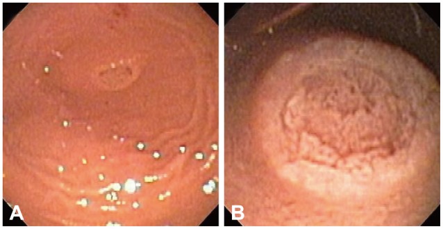
Fig. 1 Endoscopic images of congenital duodenal stenosis. (A) Duodenal bulb with an annular structure and no appreciable lumen. (B) Closer view of the villous mucosa in the center of the structure.
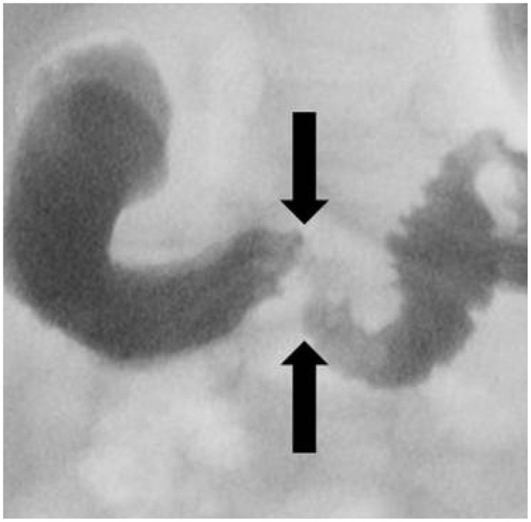
Fig. 2 Upper gastrointestinal series showing a dilated duodenal bulb and narrowing of the second portion of the duodenum (arrows), consistent with congenital duodenal stenosis.
Reference
-
1. Escobar MA, Ladd AP, Grosfeld JL, et al. Duodenal atresia and stenosis: long-term follow-up over 30 years. J Pediatr Surg. 2004; 39:867–871. PMID: 15185215.
Article2. Fabia J, Drolette M. Malformations and leukemia in children with Down's syndrome. Pediatrics. 1970; 45:60–70. PMID: 4243491.
Article3. Dalla Vecchia LK, Grosfeld JL, West KW, Rescorla FJ, Scherer LR, Engum SA. Intestinal atresia and stenosis: a 25-year experience with 277 cases. Arch Surg. 1998; 133:490–496. PMID: 9605910.4. Vinocur DN, Lee EY, Eisenberg RL. Neonatal intestinal obstruction. AJR Am J Roentgenol. 2012; 198:W1–W10. PMID: 22194504.
Article5. Grosfeld JL, Rescorla FJ. Duodenal atresia and stenosis: reassessment of treatment and outcome based on antenatal diagnosis, pathologic variance, and long-term follow-up. World J Surg. 1993; 17:301–309. PMID: 8337875.
Article6. Savino A, Rollo V, Chiarelli F. Congenital duodenal stenosis and annular pancreas: a delayed diagnosis in an adolescent patient with Down syndrome. Eur J Pediatr. 2007; 166:379–380. PMID: 17043846.
Article7. Chhabra R, Suresh BR, Weinberg G, Marion R, Brion LP. Duodenal atresia presenting as hematemesis in a premature infant with Down syndrome. Case report and review of the literature. J Perinatol. 1992; 12:25–27. PMID: 1532826.8. Sachs BF, Feldman W. Upper gastrointestinal bleeding associated with congenital duodenal stenosis and Down's syndrome. Clin Pediatr (Phila). 1973; 12:21A passim.
Article
- Full Text Links
-
- Actions
-
Cited
- CITED
-
- Close
- Share
-
- Similar articles
-
- A Case of Resolution of Duodenal Obstruction Caused by Duodenal Ulcer after the Eradication of Helicobacter pylori Infection
- A Case of Successful Management of Hypertrophic Pyloric Stenosis with Endoscopic Balloon Dilatation
- A Case of Intramural Duodenal Hematoma Complicated with Obstructive Jaundice and Pancreatitis following Endoscopic Hemostasis
- A Clinical Study of Congenital Duodenal Obstruction
- Retroperitoneal Duodenal Perforation Following a Endoscopic Sphincterotomy: A case report
