Radiation-Induced Autophagy Contributes to Cell Death and Induces Apoptosis Partly in Malignant Glioma Cells
- Affiliations
-
- 1Specific Organs Cancer Branch, National Cancer Center, Goyang, Korea.
- 2Department of Neurosurgery, University of Texas, MD Anderson Cancer Center, Houston, TX, USA.
- 3Department of Microbiology, Ajou University School of Medicine, Suwon, Korea.
- 4Cancer Cell and Molecular Biology Branch, National Cancer Center, Goyang, Korea.
- 5Neuro-oncology Clinic, National Cancer Center, Goyang, Korea. nsghs@ncc.re.kr
- KMID: 2132801
- DOI: http://doi.org/10.4143/crt.2013.159
Abstract
- PURPOSE
Radiation-induced autophagy has been shown to play two different roles, in malignant glioma (MG) cells, cytocidal or cytoprotective. However, neither the role of radiation-induced autophagy for cell death nor the existence of autophagy-induced apoptosis, a well-known cell-death pathway after irradiation, has been verified yet.
MATERIALS AND METHODS
We observed both temporal and dose-dependent response patterns of autophagy and apoptosis to radiation in MG cell lines. Additionally, we investigated the role of autophagy in apoptosis through knockdown of autophagy-related proteins.
RESULTS
Autophagic activity measured by staining of acidic vesicle organelles and Western blotting of LC-3 protein increased in proportion to radiation dose from day 1 to 5 after irradiation. Apoptosis measured by annexin-V staining and Western blotting of cleaved poly(ADP-ribose) polymerase demonstrated relatively late appearance 3 days after irradiation that increased for up to 7 days. Blocking of pan-caspase (Z-VAD-FMK) did not affect apoptosis after irradiation, but silencing of Atg5 effectively reduced radiation-induced autophagy, which decreased apoptosis significantly. Inhibition of autophagy in Atg5 knockdown cells was shown to be beneficial for cell survival. Stable transfection of GFP-LC3 cells was observed after irradiation. Annexin-V was localized in cells bearing GFP-LC3 punctuated spots, indicating autophagy in immunofluorescence. Some of these punctuated GFP-LC3 bearing cells formed conglomerated spots and died in final phase.
CONCLUSION
These findings suggest that autophagy appears earlier than apoptosis after irradiation and that a portion of the apoptotic population that appears later is autophagy-dependent. Thus, autophagy is a pathway to cell death after irradiation of MG cells.
Keyword
MeSH Terms
Figure
-
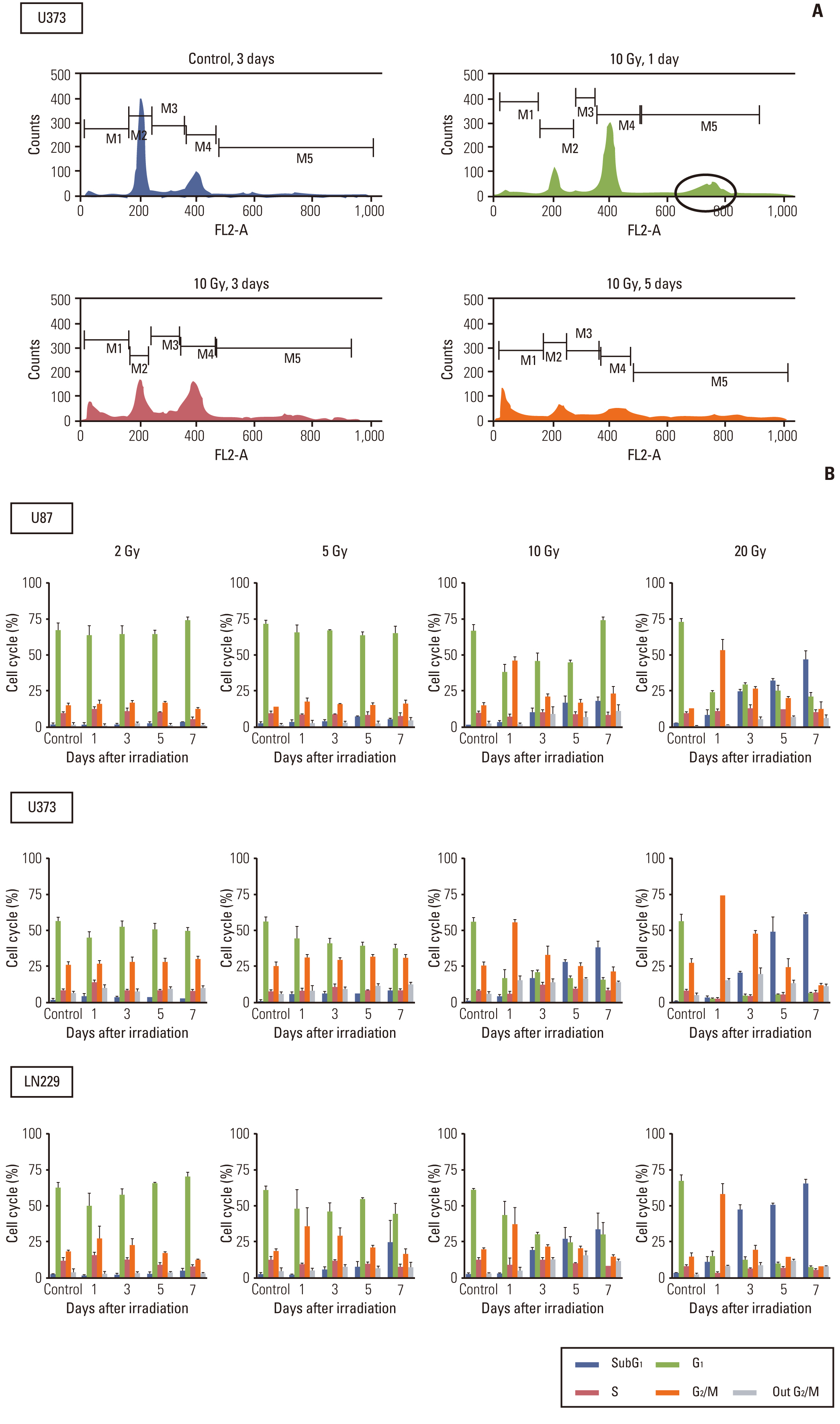
Fig. 1. Cell cycle analysis of malignant glioma cells after irradiation. Cell cycle was determined according to the DNA content in cells by flow cytometry. (A) G2/M arrest occurred as early as 1 day after irradiation (upper, right panel) and earlier than subG1 accumulations, which became apparent after 3 days (lower panels). Aneuploidy was observed at a position of 4n (circle) on 1 day after irradiation (upper, right panel). (B) Malignant glioma cells were irradiated with different doses of radiation and incubated for the indicated time. Radiation-induced G2/M arrest was minimal or recovered without a significant increase of the subG1 population in response to sublethal dose (2 Gy for all 3 cell lines and 5 Gy for U373 and U87), but prominent G2/M arrest decreased from 1 day after irradiation and was transformed into subG1 accumulation from 3 to 7 days after irradiation with lethal dose (5 Gy or more for LN229 and 10 Gy or more for U373 and U87).

Fig. 2. Analysis of autophagy after radiation according to time and radiation dose. (A) Glioma cells were irradiated with different doses of radiation and then incubated for the indicated times. To measure acidic vesicular organelle (AVO), which is indicative of autophagy formation, cells were stained with acridine orange and analyzed quantitatively by flow cytometry. Data from three independent experiments were combined. The error bars indicate ±standard error of mean. (B) Western blot analysis of LC3 according to time and radiation dose. Western blotting of LC3 generated two separate bands (an upper one of 18-kDa for LC3-bI and a lower one of 16-kDa for LC3-bII). An increase in the lower band accompanied by an increase in the ratio of the lower to the upper band is indicative of autophagy. Each membrane was stripped and re-blotted with β-actin to confirm equal loading. (C) U373 cells stably expressing GFP-LC3 revealed an increase of LC3 dots from 5 to 10 Gy and from 3 to 5 days (×200). Cells bearing GFP-LC3 puncta, which were quantified and averaged from 10 high-power fields, showed a dose-dependent increase at 3 days after irradiation (D) and an increase from 3 to 5 days after irradiation at 10 Gy (E).
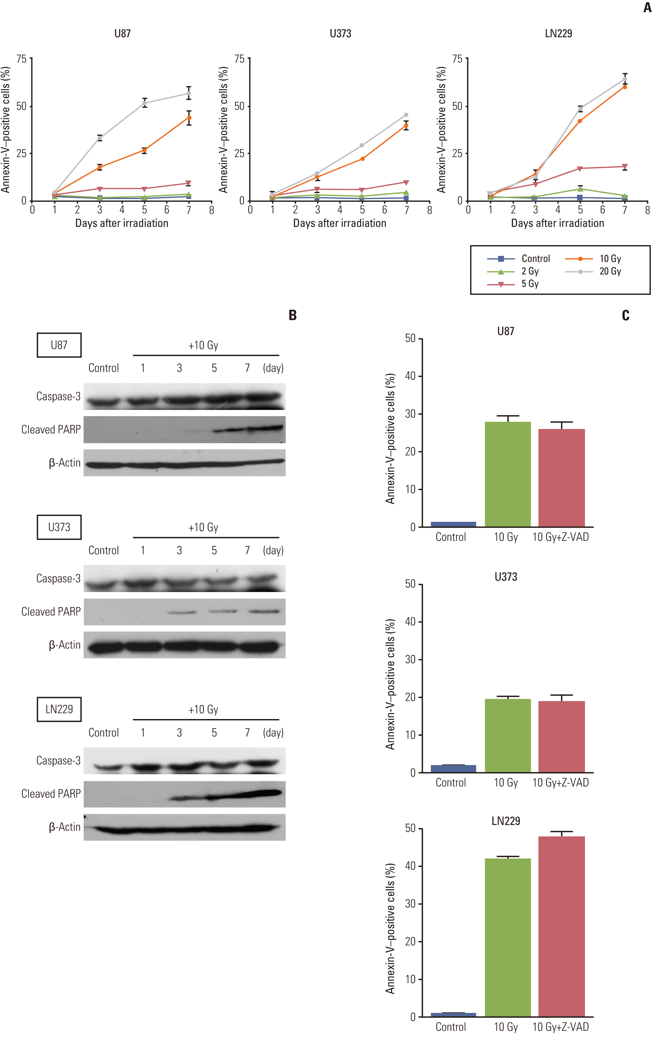
Fig. 3. Analysis of apoptosis after irradiation according to time and radiation dose. (A) Glioma cells were treated with different doses of radiation and then incubated for the indicated times, after which they were analyzed quantitatively by flow cytometry for detection of annexin-V staining, which is an early marker of apoptosis. Annexin-V–positive cells were not observed at 1 day after radiation, but appeared after 3 days and increased thereafter. Data from three independent experiments were combined. The error bars indicate ±standard error of mean.(B) Cleaved poly(ADP-ribose) polymerase (PARP), a marker of late apoptosis, was not detected at 1 day after irradiation with 10 Gy. However, it was present at 3 days after irradiation and its expression increased for up to 7 days. Caspase-3 did not change after irradiation. Each membrane was stripped and re-blotted with β-actin to confirm equal loading. (C) Cells were treated with Z-VAD-FMK (50 μM) immediately after irradiation with 10 Gy to determine if radiation-induced apoptosis in malignant glioma cells is caspase-dependent. Apoptosis was then observed and compared to that of irradiated parent cells without the inhibitor at 5 days after the irradiation.
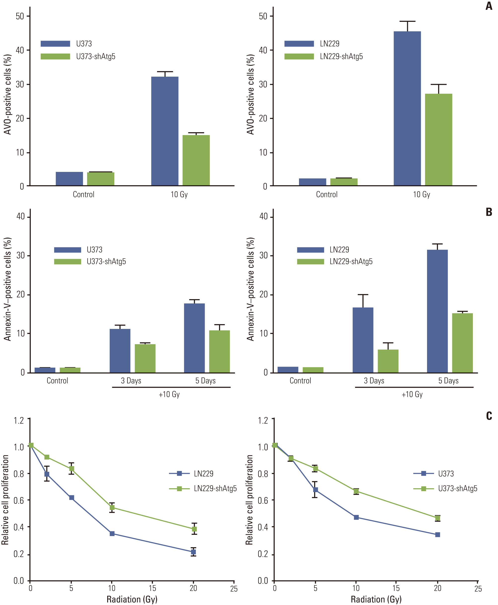
Fig. 4. Inhibition of autophagy significantly reduces apoptosis after irradiation and results in a cell survival benefit. (A) Knockdown of Atg5 effectively inhibited autophagy formation after irradiation in both U373 and LN229 cells. (B) The inhibition of autophagy translated into reduced apoptosis measured by annexin-V staining at 3 and 5 days after irradiation with 10 Gy. (C) Cell survival assay showed that autophagy inhibition resulted in cell survival. Data from three independent experiments were combined. The error bars indicate ±standard error of mean. AVO, acidic vesicular organelle.
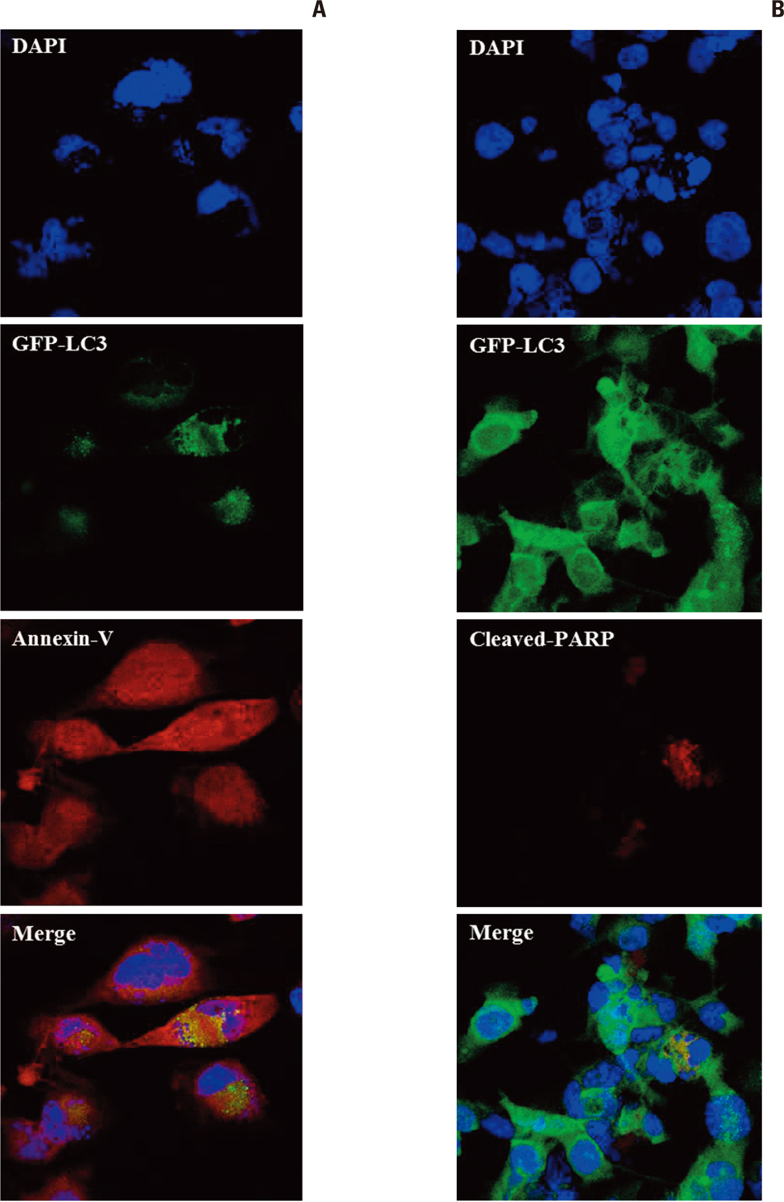
Fig. 5. Immunofluorescence of GFP-LC3, an indicator of autophagy, stably expressing U373 cells. Annexin-V and poly(ADPribose) polymerase (PARP) were used as markers of early and late apoptosis, respectively, at 5 days after irradiation with 10 Gy (×400). (A) Annexin-V is localized to cells showing a GFP-LC3 punctuated pattern representing autophagy. This co-localization suggests that autophagic cells are being converted to apoptosis. (B) Cells positive for PARP do not show a typical punctuated pattern of GFP-LC3, but show multi-nucleated cells (aneuploidy).
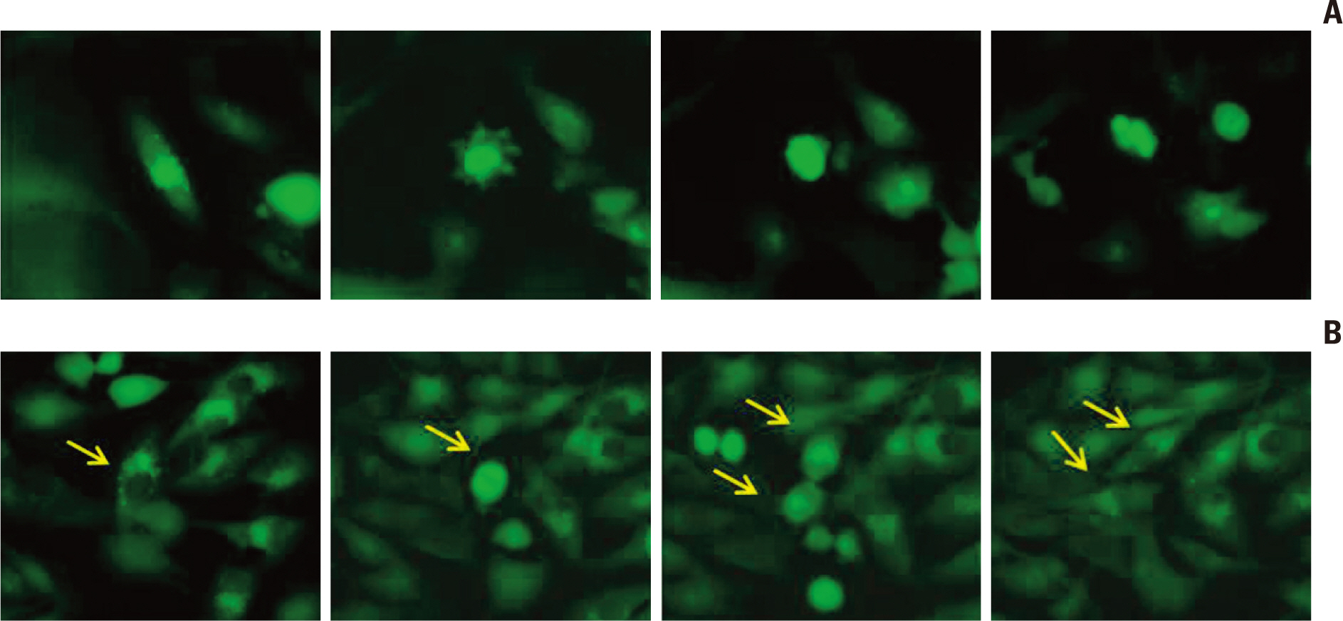
Fig. 6. Live cell image analysis of autophagic cells bearing punctuated GFP-LC3 dots (arrows) after irradiation (10 Gy) (× 200). (A) Autophagic cells showing clumping of LC3 dots and entering necrosis. (B) Other autophagic cells divide and are clear of LC3 dots.
Cited by 1 articles
-
Dexamethasone Interferes with Autophagy and Affects Cell Survival in Irradiated Malignant Glioma Cells
Alfred Komakech, Ji-Hye Im, Ho-Shin Gwak, Kyue-Yim Lee, Jong Heon Kim, Byong Chul Yoo, Heesun Cheong, Jong Bae Park, Ji Woong Kwon, Sang Hoon Shin, Heon Yoo
J Korean Neurosurg Soc. 2020;63(5):566-578. doi: 10.3340/jkns.2019.0187.
Reference
-
References
1. Walker MD, Strike TA, Sheline GE. An analysis of dose-effect relationship in the radiotherapy of malignant gliomas. Int J Radiat Oncol Biol Phys. 1979; 5:1725–31.
Article2. Hochberg FH, Pruitt A. Assumptions in the radiotherapy of glioblastoma. Neurology. 1980; 30:907–11.
Article3. Olive PL, Durand RE. Apoptosis: an indicator of radiosensitivity in vitro? Int J Radiat Biol. 1997; 71:695–707.4. Shinomiya N. New concepts in radiation-induced apoptosis: 'premitotic apoptosis' and 'postmitotic apoptosis'. J Cell Mol Med. 2001; 5:240–53.
Article5. Ning S, Knox SJ. G2/M-phase arrest and death by apoptosis of HL60 cells irradiated with exponentially decreasing low-doserate gamma radiation. Radiat Res. 1999; 151:659–69.6. Kondo Y, Kanzawa T, Sawaya R, Kondo S. The role of autophagy in cancer development and response to therapy. Nat Rev Cancer. 2005; 5:726–34.
Article7. Shintani T, Klionsky DJ. Autophagy in health and disease: a double-edged sword. Science. 2004; 306:990–5.
Article8. Lambert LA, Qiao N, Hunt KK, Lambert DH, Mills GB, Meijer L, et al. Autophagy: a novel mechanism of synergistic cytotoxicity between doxorubicin and roscovitine in a sarcoma model. Cancer Res. 2008; 68:7966–74.
Article9. Paglin S, Hollister T, Delohery T, Hackett N, McMahill M, Sphicas E, et al. A novel response of cancer cells to radiation involves autophagy and formation of acidic vesicles. Cancer Res. 2001; 61:439–44.10. Yao KC, Komata T, Kondo Y, Kanzawa T, Kondo S, Germano IM. Molecular response of human glioblastoma multiforme cells to ionizing radiation: cell cycle arrest, modulation of the expression of cyclin-dependent kinase inhibitors, and autophagy. J Neurosurg. 2003; 98:378–84.11. Ito H, Daido S, Kanzawa T, Kondo S, Kondo Y. Radiation-induced autophagy is associated with LC3 and its inhibition sensitizes malignant glioma cells. Int J Oncol. 2005; 26:1401–10.
Article12. Yousefi S, Perozzo R, Schmid I, Ziemiecki A, Schaffner T, Scapozza L, et al. Calpain-mediated cleavage of Atg5 switches autophagy to apoptosis. Nat Cell Biol. 2006; 8:1124–32.
Article13. Luo S, Rubinsztein DC. Atg5 and Bcl-2 provide novel insights into the interplay between apoptosis and autophagy. Cell Death Differ. 2007; 14:1247–50.
Article14. Kanzawa T, Germano IM, Komata T, Ito H, Kondo Y, Kondo S. Role of autophagy in temozolomide-induced cytotoxicity for malignant glioma cells. Cell Death Differ. 2004; 11:448–57.
Article15. Fowler JF. The linear-quadratic formula and progress in fractionated radiotherapy. Br J Radiol. 1989; 62:679–94.
Article16. Shinomiya N, Kuno Y, Yamamoto F, Fukasawa M, Okumura A, Uefuji M, et al. Different mechanisms between premitotic apoptosis and postmitotic apoptosis in X-irradiated U937 cells. Int J Radiat Oncol Biol Phys. 2000; 47:767–77.
Article17. Su LN, Little JB. Prolonged cell cycle delay in radioresistant human cell lines transfected with activated ras oncogene and/or simian virus 40 T-antigen. Radiat Res. 1993; 133:73–9.
Article18. Stuschke M, Sak A, Wurm R, Sinn B, Wolf G, Stuben G, et al. Radiation-induced apoptosis in human non-small-cell lung cancer cell lines is secondary to cell-cycle progression beyond the G2-phase checkpoint. Int J Radiat Biol. 2002; 78:807–19.
Article19. Broker LE, Huisman C, Ferreira CG, Rodriguez JA, Kruyt FA, Giaccone G. Late activation of apoptotic pathways plays a negligible role in mediating the cytotoxic effects of discodermolide and epothilone B in non-small cell lung cancer cells. Cancer Res. 2002; 62:4081–8.20. Cummings BS, Kinsey GR, Bolchoz LJ, Schnellmann RG. Identification of caspase-independent apoptosis in epithelial and cancer cells. J Pharmacol Exp Ther. 2004; 310:126–34.
Article21. Chaitanya GV, Babu PP. Differential PARP cleavage: an indication of heterogeneous forms of cell death and involvement of multiple proteases in the infarct of focal cerebral ischemia in rat. Cell Mol Neurobiol. 2009; 29:563–73.
Article22. Gwak HS, Shingu T, Chumbalkar V, Hwang YH, DeJournett R, Latha K, et al. Combined action of the dinuclear platinum compound BBR3610 with the PI3-K inhibitor PX-866 in glioblastoma. Int J Cancer. 2011; 128:787–96.
Article23. Codogno P, Meijer AJ. Atg5: more than an autophagy factor. Nat Cell Biol. 2006; 8:1045–7.
Article24. Shimizu S, Kanaseki T, Mizushima N, Mizuta T, Arakawa-Kobayashi S, Thompson CB, et al. Role of Bcl-2 family proteins in a non-apoptotic programmed cell death dependent on autophagy genes. Nat Cell Biol. 2004; 6:1221–8.
Article25. Kim KW, Mutter RW, Cao C, Albert JM, Freeman M, Hallahan DE, et al. Autophagy for cancer therapy through inhibition of pro-apoptotic proteins and mammalian target of rapamycin signaling. J Biol Chem. 2006; 281:36883–90.
Article
- Full Text Links
-
- Actions
-
Cited
- CITED
-
- Close
- Share
-
- Similar articles
-
- Dexamethasone Interferes with Autophagy and Affects Cell Survival in Irradiated Malignant Glioma Cells
- Memantine Induces NMDAR1-Mediated Autophagic Cell Death in Malignant Glioma Cells
- Regulatory Role of Autophagy in Globular Adiponectin-Induced Apoptosis in Cancer Cells
- Induction of Apoptosis and Autophagy in UVB-Treated HaCaT Cells
- The Impact of Autophagy on the Cigarette Smoke Extract-Induced Apoptosis of Bronchial Epithelial Cells
