Ancient Schwannoma at the Olfactory Groove Mimicking Meningioma: A Case Report
- Affiliations
-
- 1Department of Radiology, Busan Paik Hospital, Inje University College of Medicine, Busan, Korea. hwjeong2000@lycos.co.kr
- KMID: 2130949
- DOI: http://doi.org/10.3348/jksr.2015.73.6.413
Abstract
- Schwannomas are benign slow-growing nerve sheath tumors, which can develop in any peripheral or central nerve that contains Schwann cells. Schwannomas located near the olfactory groove are extremely rare and radiological diagnosis can be difficult. Moreover, ancient schwannoma is an uncommon variant, and radiologic findings are rarely reported. Herein, we reported a surgically confirmed case of ancient schwannoma at the olfactory groove in a 44-year-old woman presenting with headache and visual disturbance. Brain magnetic resonance imaging (MRI) showed a solid and cystic extra-axial mass located in the subfrontal area mimicking an olfactory groove meningioma. Histopathologic diagnosis of ancient schwannoma was confirmed by immunohistochemical staining for S100, CD56, vimentin, and other markers. Furthermore, we described the clinical manifestations, MRI characteristics, and histopathologic findings of the case, and presented a review of related literature.
MeSH Terms
Figure
-
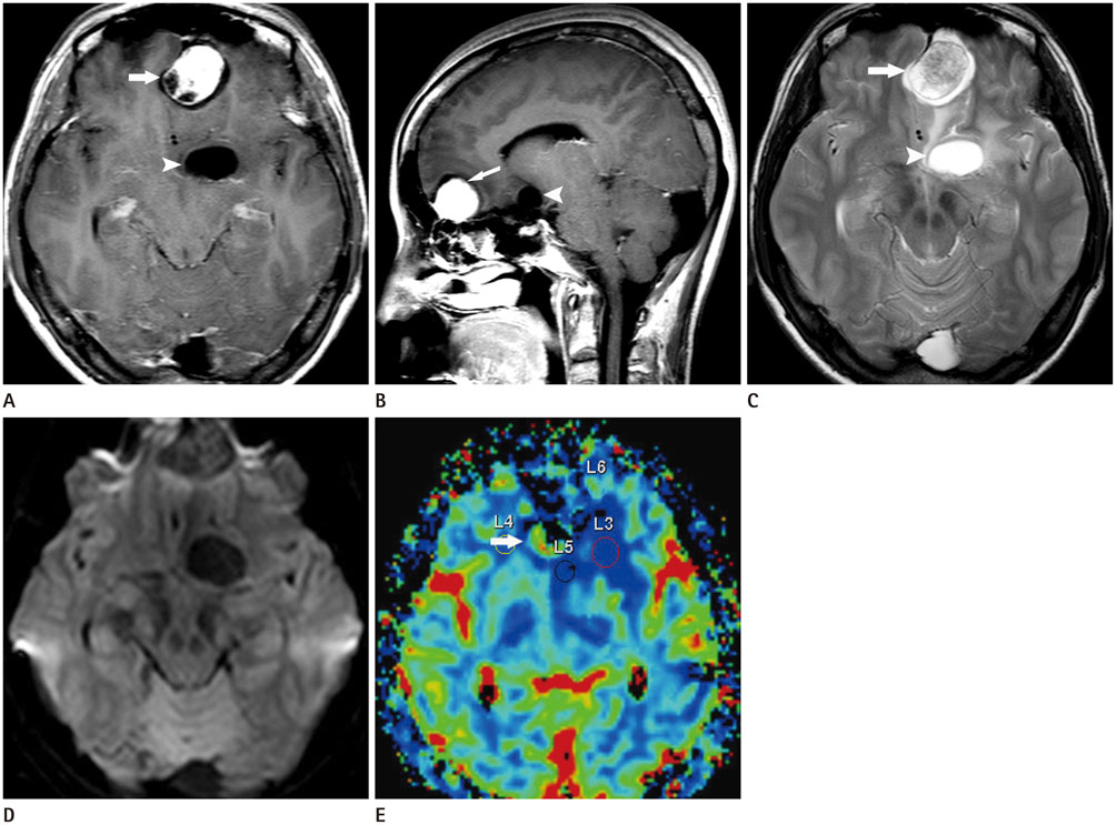
Fig. 1 A 44-year-old woman with headache and visual disturbance. A, B. Contrast-enhanced T1-weighted axial (A) and sagittal (B) magnetic resonance (MR) images, showing a heterogeneously enhanced extra-axial mass with cystic areas in the left subfrontal area. Solid enhancement (arrow) in the anterior portion and a non-enhanced cystic area (arrowhead) in the posterior portion are noted. The lesion shows a clear margin with the surrounding tissue. C. T2-weighted axial MR image showing a solid mass (arrow) surrounded by high signal intensity, implying a cystic component (arrowhead). D. Diffusion MR image showing no definite diffusion restriction in the solid portion. E. Perfusion MR image showing increased relative cerebral blood volume in the solid portion (arrow) and no definite change in the cystic portion.

Fig. 2 Histologic examination, showing relatively compact areas of spindle cells (Antoni A pattern) (A), Verocay bodies are formed by palisade Schwann cells (arrows) (magnification × 40) (B). Extensive vascular hyalinization (arrowheads), one of the degenerative changes of ancient schwannoma is seen (magnification × 10). Diffuse staining for CD56 (C) and S100 (D) in cell nuclei and cytoplasm, which is typically more prominent in hypercellular areas (Antoni A) than in hypocellular areas (Antoni B), is noted (magnification × 40).
Reference
-
1. Park JH, Kim TY, Park JT, Kim JM. Olfactory groove schwannoma. J Korean Neurosurg Soc. 2006; 39:156–158.2. Urich H, Tien RD. Tumors of the cranial, spinal and peripheral nerve sheaths. In : Bigner DD, McLendon RE, Bruner JM, Russell DS, editors. Russell and Rubinstein's pathology of tumours of the nervous system. London, New York: Arnold, Oxford University Press;1988. p. 141–193.3. Lee YS, Kim JO, Park SE. Ancient schwannoma of the thigh mimicking a malignant tumour: a report of two cases, with emphasis on MRI findings. Br J Radiol. 2010; 83:e154–e157.4. Choi YS, Sung KS, Song YJ, Kim HD. Olfactory schwannoma-case report. J Korean Neurosurg Soc. 2009; 45:103–106.5. Isobe K, Shimizu T, Akahane T, Kato H. Imaging of ancient schwannoma. AJR Am J Roentgenol. 2004; 183:331–336.6. Adachi K, Yoshida K, Miwa T, Ikeda E, Kawase T. Olfactory schwannoma. Acta Neurochir (Wien). 2007; 149:605–610. discussion 610-611.7. Santhosh K, Kesavadas C, Radhakrishnan VV, Thomas B, Kapilamoorthy TR, Gupta AK. Usefulness of T2*-weighted MR sequence for the diagnosis of subfrontal schwannoma. J Neuroradiol. 2007; 34:330–333.8. Timothy J, Chakrabarty A, Rice A, Marks P. Olfactory groove schwannoma revisited. Acta Neurochir (Wien). 1999; 141:671–672.9. Zagardo MT, Castellani RJ, Rees JH, Rothman MI, Zoarski GH. Radiologic and pathologic findings of intracerebral schwannoma. AJNR Am J Neuroradiol. 1998; 19:1290–1293.10. Luo B, Sun G, Zhang B, Liang K, Wen J, Fang K. Neuroradiological findings of intracranial schwannomas not arising from the stems of cranial nerves. Br J Radiol. 2004; 77:1016–1021.
