Epiphyseal Hemangioma of the Humeral Head: Imaging Findings and Literature Review
- Affiliations
-
- 1Department of Radiology, Kyung Hee University Hospital at Gangdong, College of Medicine, Kyung Hee University, Seoul, Korea. jinooki@daum.net
- 2Department of Orthopaedic Surgery, Kyung Hee University Hospital at Gangdong, College of Medicine, Kyung Hee University, Seoul, Korea.
- 3Department of Pathology, Kyung Hee University Hospital at Gangdong, College of Medicine, Kyung Hee University, Seoul, Korea.
- 4Department of Radiology, Kyung Hee University Hospital, College of Medicine, Kyung Hee University, Seoul, Korea.
- KMID: 2130947
- DOI: http://doi.org/10.3348/jksr.2015.73.6.403
Abstract
- We describe a case of an epiphyseal hemangioma in the humeral head of a 20-year-old man. On plain radiographs, the lesion showed no gross abnormality. The computed tomography images demonstrated the presence of an irregular and lobulating osteolytic lesion with a peripheral sclerotic rim and focal cortical defects. The magnetic resonance images showed an ill-defined low signal intensity on T1-weighted images and mixed low and high signal intensities on T2-weighted images. Additionally, ill-defined marrow enhancement with inner low signal lines was noted in this lesion. The patient was treated with curettage and a bone chip graft. The present case is instructive in the differential diagnosis of epiphyseal bone tumors; furthermore, the possibility of an intraosseous hemangioma should also be considered.
MeSH Terms
Figure
-

Fig. 1 An anteroposterior radiograph of the right shoulder shows no gross abnormalities in the humeral head.

Fig. 2 CT scans show an irregular and lobulating osteolytic lesion with a peripheral sclerotic rim and thickened internal trabeculae (arrow). On coronal reformatted images, a focal cortical defect (arrow) is suggested.
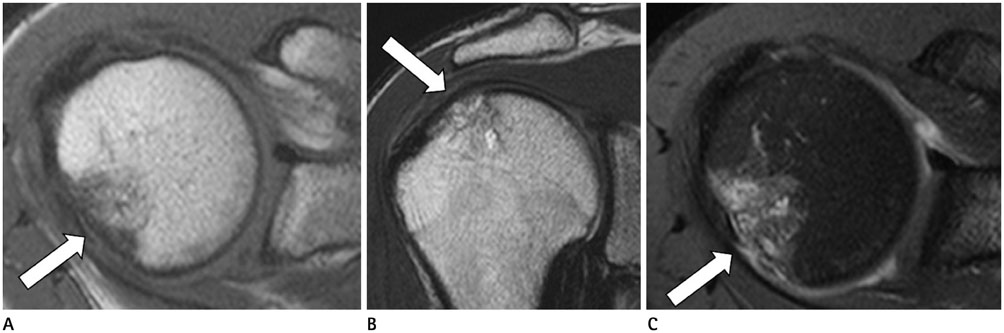
Fig. 3 Magnetic resonance images show irregular low signal intensity on T1-weighted images (A, arrow; T1WI; axial plane; TR/TE, 600/20 ms), mixed low and high signal intensities on T2-weighted images (B, arrow; T2WI; coronal plane; TR/TE, 4400/80 ms) and inhomogeneous enhancement on gadolinium-enhanced fat-suppressed T1WI (C, arrow; CE-FS T1WI; axial plane; TR/TE, 670/20 ms). In addition, inner and peripheral low signal lines in this lesion are demonstrated on all MR sequences. CE-FS = contrast-enhanced fat-suppressed, TE = echo time, TR = repetition time

Fig. 4 A photomicrograph (A) of an intraosseous hemangioma reveals numerous vascular channels (arrows) of various sizes and shapes. The irregular architecture of the thickened bony trabeculae (T) is noted (hematoxylin and eosin staining, original magnification, × 40). Immunostaining for factor VIII (B) shows vascular channels of a variety of shapes and sizes (arrows), lined by endothelial cells (polymer method; original magnification, × 200).
Reference
-
1. Unni KK. Benign vascular tumors. In : Unni KK, Inwards CY, editors. Dahlin's Bone Tumors: General Aspects and Data on 10,165 Cases. Philadelphia: Lippincott Williams & Wilkins;2010. p. 262–271.2. Resnick D. Tumors of vascular differentiation. In : Resnick D, editor. Diagnosis of Bone and Joint Disorders. Philadelphia: Saunders;1995. p. 3821–3846.3. Boumdin H, Rachid K, Mahi M, Chaouir S, Benameur M. [Hemangioma of the humerus: value of imaging]. J Radiol. 2002; 83(9 Pt 1):1088–1089.4. Yamamoto T, Kurosaka M, Mizuno K. Juxta-articular hemangioma of long bone. Skeletal Radiol. 2000; 29:535–537.5. Mirra JM. Vascular tumors. In : Mirra JM, Picci P, Gold RH, editors. Bone Tumors: Clinical, Radiologic, and Pathologic Correlations. Philadelphia: Lea & Febiger;1989. p. 1338–1478.6. Pandey S, Pandey AK. Osseous haemangiomas. Arch Orthop Trauma Surg. 1981; 99:23–28.7. Dorfman HD, Czerniak B. Vascular lesions. In : Dorfman HD, Czerniak B, editors. Bone Tumors. St. Louis: Mosby;1998. p. 729–814.8. Ross JS, Masaryk TJ, Modic MT, Carter JR, Mapstone T, Dengel FH. Vertebral hemangiomas: MR imaging. Radiology. 1987; 165:165–169.9. Murphey MD, Fairbairn KJ, Parman LM, Baxter KG, Parsa MB, Smith WS. From the archives of the AFIP. Musculoskeletal angiomatous lesions: radiologic-pathologic correlation. Radiographics. 1995; 15:893–917.10. Matsumoto K, Ishizawa M, Okabe H, Taniguchi I. Hemangioma of bone arising in the ulna: imaging findings with emphasis on MR. Skeletal Radiol. 2000; 29:231–234.
- Full Text Links
-
- Actions
-
Cited
- CITED
-
- Close
- Share
-
- Similar articles
-
- Reconstruction with Vascularized Fibular Epiphyseal Transplantation of Humeral Head Deformity by Septic Arthritis
- Cavernous Hemangioma of the External Auditory Canal: A Case Report
- Cavernous Hemangioma of the Masseter Muscle
- A Case of Subcutaneous Cavernous Hemangioma Presenting as a Nasolabial Fold Mass
- Rapid progressive atypical atraumatic osteonecrosis of humeral head: a case report
