J Korean Soc Radiol.
2015 Dec;73(6):389-392. 10.3348/jksr.2015.73.6.389.
A Rare Case of Cystic Subepithelial Tumor in the Stomach: Gastric Adenomyoma
- Affiliations
-
- 1Department of Radiology, Kyungpook National University Hospital, Daegu, Korea. yjjang@knu.ac.kr
- 2Department of Internal Medicine, Kyungpook National University Hospital, Daegu, Korea.
- KMID: 2130944
- DOI: http://doi.org/10.3348/jksr.2015.73.6.389
Abstract
- Gastric adenomyoma is a rare benign subepithelial tumor, characteristically composed of mucosal structures and a prominent smooth muscle stroma. Because of rarity and the nonspecific computed tomography (CT) features, it is difficult to diagnose gastric adenomyoma before operation. In our case, gastric adenomyoma showed a well-circumscribed cystic subepithelial mass with uneven wall thickness on a CT scan, similar to the findings of former reports. The radiologic differential diagnosis can be narrowed down to several diseases, including duplication cysts, gastritis cystica profunda, brunner's gland hyperplasia and solid tumors with cystic degeneration. Also, adenomyoma could be included in the differential diagnosis of gastric cystic subepithelial masses, especially in the distal part of the stomach.
MeSH Terms
Figure
-
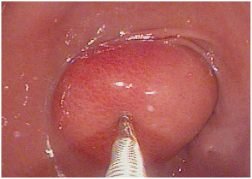
Fig. 1 An upper gastrointestinal endoscopic examination shows a round mass protruding in to the lumen in the anterior wall of the gastric antrum. Relatively normal overlying mucosa is observed, and the mass is smoothly compressible by an endoscopic forceps.
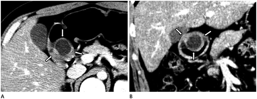
Fig. 2 Axial (A) and coronal (B) reformatted contrast-enhanced CT images show a well-circumscribed cystic mass (arrows), with uneven wall thickening, in the gastric antrum.
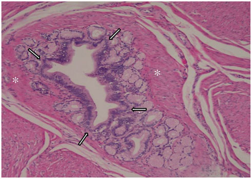
Fig. 3 A microscopy of the specimen shows epithelial elements (arrows) in the form of individual glandular structures supported by the hypertrophic smooth muscle fibers (*) of the stroma (hematoxylin & eosin stain, × 50).
Reference
-
1. Erberich H, Handt S, Mittermayer C, Tietze L. Simultaneous appearance of an adenomyoma and pancreatic heterotopia of the stomach. Virchows Arch. 2000; 436:172–174.2. Zhu HN, Yu JP, Luo J, Jiang YH, Li JQ, Sun WY. Gastric adenomyoma presenting as melena: a case report and literature review. World J Gastroenterol. 2010; 16:1934–1936.3. Chapple CR, Muller S, Newman J. Gastric adenocarcinoma associated with adenomyoma of the stomach. Postgrad Med J. 1988; 64:801–803.4. Rhim JH, Kim WS, Choi YH, Cheon JE, Park SH. Radiological findings of gastric adenomyoma in a neonate presenting with gastric outlet obstruction. Pediatr Radiol. 2013; 43:628–630.5. Takeyama J, Sato T, Tanaka H, Nio M. Adenomyoma of the stomach mimicking infantile hypertrophic pyloric stenosis. J Pediatr Surg. 2007; 42:E11–E12.6. Min SH, Kim HY, Kim SH, Jung SE, Park KW, Kim WS, et al. Gastric adenomyoma mimicking gastric duplication cyst in a 5-year-old girl. J Pediatr Surg. 2012; 47:1019–1022.7. Yoon KH, Eun DY, Kim JH, Lee SO, Kim HS, Lee DW. Gastric adenomyoma in the stomach body: a case report. J Med Case Rep. 2014; 8:385.8. Matsushita M, Takakuwa H, Nishio A. Endosonographic features of gastric adenomyoma, a type of ectopic pancreas. Endoscopy. 2003; 35:621–622. author reply 6239. Kagawa S, Fujiwara T, Nishizaki M, Naomoto Y, Hiroshi I, Tanaka N. Adenomyoma of the stomach presenting as localized peritonitis. Dig Dis Sci. 2007; 52:3184–3187.
- Full Text Links
-
- Actions
-
Cited
- CITED
-
- Close
- Share
-
- Similar articles
-
- Incidental Adenomyoma of Stomach Associated with Early Gastric Cancer
- Adenomyoma in the Body of Stomach Presenting as a Pedunculated Polyp Treated by Endoscopic Mucosal Resection
- A case of huge extrauterine endometrioid-type adenomyoma with cystic change: A case report and literature review
- A Case of Cystic Degeneration of Uterine Adenomyosis
- A case of cystic adenomyoma of the uterus after complete abortion without transcervical curettage
