Synthetic CDCA Derivatives-Induced Apoptosis of Stomach Cancer Cell Line SNU-1 Cells
- Affiliations
-
- 1Department of Family Medicine, Dong-A University College of Medicine, Busan, Korea. jspark@daunet.donga.ac.kr
- 2Department of Surgery, Dong-A University College of Medicine, Busan, Korea.
Abstract
- PURPOSE
This study was conducted to explore whether CDCA derivatives induce apoptosis in a stomach cancer cell line, and to dissect the detailed mechanism underlying apoptosis. MATERIALS AND METHODS: The human stomach cancer cell line, SNU-1, cells were treated with the synthetic CDCA derivatives, HS-1199 and HS-1200. DNA and mitochondrial stains were used to detect apoptotic cells by fluorescence imaging or flow cytometry. The caspase-3 activity was measured by Western blotting. RESULTS: Both the HS-1199 and HS-1200 induced decreased viabilities of the SNU-1 cells, in time-dependent manners. The CDCA derivatives demonstrated various apoptosis hallmarks, such as mitochondrial changes reduction of MMP, cytochrome c release, and Smac/ DIABLO translocation), activation of caspase-3 (resulting in the degradation of PARP and DFF45), DNA fragmentation and nuclear condensation. CONCLUSION: The CDCA derivatives, HS-1199 and HS- 1200, both induced apoptosis of the SNU-1 gastric cancer cells in caspase- and mitochondria-dependent fashions. Many important issues relating to their therapeutic applications remain to be elucidated.
MeSH Terms
Figure
-
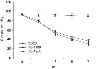
Fig. 1 The viability of SNU-1 was decreased after the CDCA derivatives treatment. 50µM of both HS-1199 and HS-1200 produced significant time-dependent decreases in the cell viability (0~7 h, p<0.01), whereas CDCA did not.
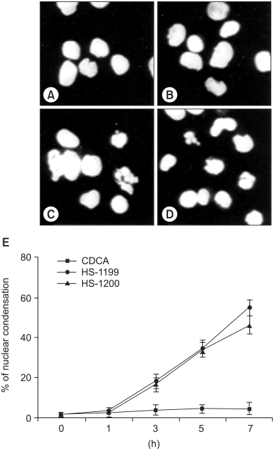
Fig. 2 Nuclear condensation induced by CDCA derivatives was demonstrated five hours after treatment. Hoechst staining demonstrated that CDCA derivatives induced a change in the nuclear morphology. Compared to the typical round nuclei of the control or CDCA-treated cells (A&B), the cells treated with 50M of both HS-1199 and HS-1200 displayed condensed and fragmented nuclei (C&D). The percentage of dead or dying cells, as determined by nuclear morphology, was significantly increased in the CDCA derivatives-treated cells in a time-dependent manner (E; 0~7 h, p<0.01).
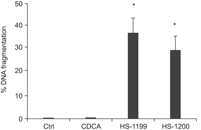
Fig. 3 CDCA derivatives produced DNA fragmentation five hours after treatment. Treatment of SNU-1 cells with 50µM of the CDCA derivatives, HS-1199 and HS-1200, resulted in DNA fragmentation, as determined by the TUNEL assay (*, p<0.01).
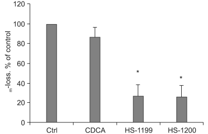
Fig. 4 CDCA derivatives produced reductions in the MMP. Loss of mitochondrial membrane potential (ΔΨm) is known to be a common event in many pathways of apoptosis induction. In this study, the potential-sensitive fluorescent probe JC-1 was employed to detect loss of ΔΨm. As depicted in figure 4, the membrane potential was rapidly reduced 5 h after treatment with 50µM CDCA derivatives. ΔΨm decreased significantly in the CDCA derivative-treated cells (*, p<0.01).
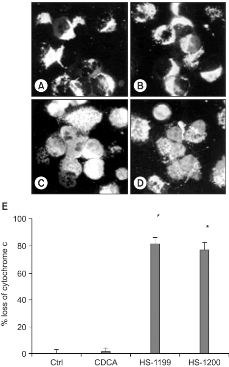
Fig. 5 CDCA derivatives produced cytochrome c release to cytosol. In the immunofluorescent study, the cytochrome c in the control and CDCA-treated cells were found in punctate patterns, in keeping with its normal mitochondrial location (A&B). The location of the cytochrome c in the mitochondria was confirmed by double staining of the HSP-60 in the same sample (data not shown). However, 50µM of the CDCA derivatives, HS-1199 and HS-1200, treatment led to the release of cytochrome c from the mitochondria into the cytosol (C&D). Quantification data (5 h) are also shown (E; *, p<0.01).
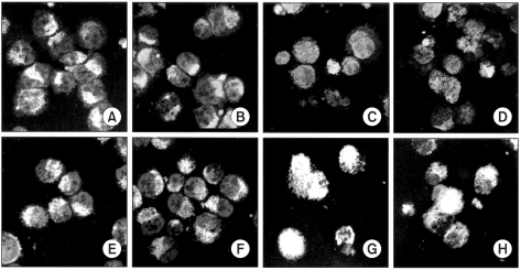
Fig. 6 AIF and Smac/DIABLO were released from mitochondria. In the immunofluorescent study, the AIF and Smac/DIABLO in the control and 50µM CDCA-treated cells were found in punctate patterns (A&B, E&F). The CDCA derivatives led to the release of both factors. The AIF was translocated onto the nucleus, whereas the Smac/DIABLO to the cytosol (C&D, G&H).
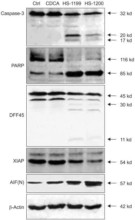
Fig. 7 Western blotting shows changes of the apoptosis-related proteins in this type of apoptosis. In the 50µM CDCA derivatives-treated cells the AIF expression increased, whereas that of the XIAP decreased. However, the CDCA derivatives showed no difference in the expression levels compared to the control or CDCA-treated cells. CDCA derivatives treatment evidently displayed activation of caspases-3, resulting in the degradations of PARP and DFF45. A 32 kDa precursor of caspase-3 was degraded, resulting in 20- and 17-kDa cleavage products. The PARP 85-kDa and DFF45 11-kDa cleavage products were shown to increase in time dependent manners by CDCA derivatives-induced apoptosis.

Fig. 8 The CDCA derivatives produced no specific cell cycle phase arrest. In the 50µM CDCA derivatives-treated cells, the percentage of cells showing DNA hypoploidy increased. However, arrest of specific cell cycle phase was not observed.
Reference
-
1. Martinez JD, Stratagoules ED, LaRue JM, Powell AA, Gause PR, Craven MT, Payne CM, Powell MB, Gerner EW, Earnest DL. Different bile acids exhibit distinct biological effects: the tumor promoter deoxycholic acid induces apoptosis and the chemopreventive agent ursodeoxycholic acid inhibits cell proliferation. Nutr Cancer. 1998; 31:111–118. PMID: 9770722.
Article2. Rust C, Karnitz LM, Paya CV, Moscat J, Simari RD, Gores GJ. The bile acid taurochenodeoxycholate activates a phosphatidylinositol 3-kinase-dependent survival signaling cascade. J Biol Chem. 2000; 275:20210–20216. PMID: 10770953.
Article3. Chu SH, Park WM, Lee KE, Pae YS. Glycochenodeoxycholic acid induces cell death in primary cultured rat hepatocyte: Apoptosis and necrosis. Korean J Physiol Pharmacol. 1999; 3:565–570.4. Powell AA, LaRue JM, Batta AK, Martinez JD. Bile acid hydrophobicity is correlated with induction of apoptosis and/or growth arrest in HCT116 cells. Biochem J. 2001; 356:481–486. PMID: 11368775.
Article5. Loogna P, Franzen L, Sipponen P, Domellof L. Effects of Helicobacter pylori and bile on N-methyl-N'-nitro-N'-nitrosoguanidine exposed antral mucosa of C57BU6 mice. Virchows Arch. 2001a; 439:661–667. PMID: 11764387.6. Loogna P, Franzen L, Sipponen P, Domellof L. Helicobacter pylori, N-methyl-N'-nitro-N'-nitrosoguanidine, and bile modulate gastric cell kinetics in experimental cancer. Virchows Arch. 2001b; 439:653–660. PMID: 11764386.
Article7. Brady LM, Beno DWA, Davis BH. Bile acid stimulation of early growth response gene and mitogen-activated protein kinase is protein kinase C-dependent. Biochem J. 1996; 316:765–769. PMID: 8670150.
Article8. LaRue JM, Stratagoules ED, Martinez JD. Deoxycholic acid-induced apoptosis is switched to necrosis by bcl-2 or calphostin C. Cancer Lett. 2000; 152:107–113. PMID: 10754212.9. Glinghammar B, Holmberg K, Rafter J. Effects of colonic luminal components on AP-1-dependent gene transcription in cultured human colon carcinoma cells. Carcinogenesis. 1999; 20:969–976. PMID: 10357775.10. Zang F, Subbaramaiah K, Altorki N, Dannenberg AJ. Dihydroxy bile acid activates the transcription of cyclooxygenase-2. J Biol Chem. 1998; 273:2424–2428. PMID: 9442092.11. Di Toro R, Campana G, Murari G, Spampinato S. Effects of specific bile acids on c-fos messenger RNA levels in human colon carcinoma Caco-2 cells. Eur J Pharm Sci. 2000; 11:291–298. PMID: 11033072.12. Choi YH, Im EO, Suh H, Jin Y, Lee WH, Yoo YH, Kim KW, Kim ND. Apoptotic activity of novel bile acid derivatives in human leukemic T cells through the activation of caspases. Int J Oncol. 2001; 18:979–984. PMID: 11295044.
Article13. Im EO, Choi YH, Paik KJ, Suh H, Jin Y, Kim KW, Yoo YH, Kim ND. Novel bile acid derivatives induce apoptosis via a p53-independent pathway in human breast carcinoma cells. Cancer Lett. 2001; 163:83–93. PMID: 11163111.
Article14. Im EO, Lee S, Suh H, Kim KW, Bae YT, Kim ND. A novel ursodeoxycholic acid derivative induces apoptosis in human MCF-7 breast cancer cells. Pharm Pharmacol Commun. 1999; 5:1–6.
Article15. Huang P, Ballal K, Plunkett W. Biochemical characterization of the protein activity responsible for high molecular weight DNA fragmentation during drug-induced apoptosis. Cancer Res. 1997; 57:3407–3414. PMID: 9270006.16. Neamati N, Fernandez A, Wright S, Kiefer J, McConkey DJ. Degradation of lamin B1 precedes oligonucleosomal DNA fragmentation in apoptotic thymocytes and isolated thymocyte nuclei. J Immunol. 1995; 154:3788–3795. PMID: 7535814.17. Konopleva M, Zhao S, Xie Z, Segall H, Younes A, Claxton DF, Estrov Z, Kornblau SM, Andreeff M. Apoptosis. Molecules and mechanisms. Adv Exp Med Biol. 1999; 457:217–236. PMID: 10500797.18. Lazebnik YA, Kaufmann SH, Desnoyers S, Poirier GG, Earnshaw WC. Cleavage of poly (ADP-ribose) polymerase by a proteinase with properties like ICE. Nature. 1994; 371:346–347. PMID: 8090205.19. Findley HW, Gu L, Yeager AM, Zhou M. Expression and regulation of Bcl-2, Bcl-xl, and Bax correlate with p53 status and sensitivity to apoptosis in childhood acute lymphoblastic leukemia. Blood. 1997; 89:2986–2993. PMID: 9108419.20. Salomons GS, Brady HJ, Verwijs-Janssen M, Van Den Berg JD, Hart AA, Behrendt H, Hahlen K, Smets LA. The Bax alpha: Bcl-2 ratio modulates the response to dexamethasone in leukaemic cells and is highly variable in childhood acute leukaemia. Int J Cancer. 1997; 71:959–965. PMID: 9185697.21. Susin SA, Lorenzo HK, Zamzami N, Marzo I, Snow BE, Brothers GM, Mangion J, Jacotot E, Costantini P, Loeffler M, Larochette N, Goodlett DR, Aebersold R, Siderovski DP, Penninger JM, Kroemer G. Molecular characterization of mitochondrial apoptosis-inducing factor. Nature. 1999; 397:441–446. PMID: 9989411.
Article22. Yerushalmi B, Dahl R, Devereaux MW, Gumpricht E, Sokol RJ. Bile acid-induced rat hepatocyte apoptosis is inhibited by antioxidants and blockers of the mitochondrial permeability transition. Hepatology. 2001; 33:616–626. PMID: 11230742.
Article23. Kroemer G, Dallaporta B, Resche-Rigon M. The mitochondrial death/life regulator in apoptosis and necrosis. Annu Rev Physiol. 1998; 60:619–642. PMID: 9558479.
Article24. Bidere N, Senik A. Caspase-independent apoptotic pathways in T lymphocytes: A minireview. Apoptosis. 2001; 6:371–375. PMID: 11483861.25. Du C, Fang M, Li L, Wang W. Smac, a mitochondrial protein that promotes cytochrome c-dependent caspase activation by eliminating IAP inhibition. Cell. 2000; 102:33–42. PMID: 10929711.
- Full Text Links
-
- Actions
-
Cited
- CITED
-
- Close
- Share
-
- Similar articles
-
- Synthetic Chenodeoxycholic Acid Derivative HS-1200-induced Apoptosis of Human Melanoma Cells
- Synthetic Bile Acid Derivative HS-1200-induced Apoptosis of Human Osteosarcoma Cells
- A Synthetic Chenodeoxycholic Acid Derivative, HS-1200-induced Apoptosis of RBL-2H3 Cells
- Modulation of Apoptosis and Cell Cycle by Synthetic Bile Acid Derivatives
- Apoptotic Effect of Co-treatment with Chios Gum Mastic and HS-1200 on G361 Human Melanoma Cell Line
