A Case of Conjunctival Lymphangioma With Clinical Manifestations of Superior Limbic Keratoconjunctivitis After Upper Lid Blepharoplasty
- Affiliations
-
- 1Department of Ophthalmology, KyungHee University School of Medicine, Seoul, Korea. khjinmd@khmc.or.kr
- 2Department of Ophthalmology, Kangwon University School of Medicine, Chuncheon, Korea.
- KMID: 2122297
- DOI: http://doi.org/10.3341/jkos.2010.51.9.1276
Abstract
- PURPOSE
To report a case of conjunctival lymphangioma with clinical manifestations of superior limbic keratoconjunctivitis after upper lid blepharoplasty.
CASE SUMMARY
A 55-year-old woman who had upper lid blepharoplasty performed 1 year before complained of pain, injection, and blurred vision in her right eye, which lasted for 2 days. The slit lamp examination revealed a poorly demarcated hypertrophic lesion with central erosion on the superior bulbar conjunctiva and a diffuse corneal erosion and edema in the area of the superior limbus. Tenderness of the superior bulbar conjunctiva and corneal erosion were treated with artificial tears, antibiotic eye drops, and a therapeutic contact lens. Slight anterior chamber reaction was found and treated by steroid eye drops; however, there was no improvement. After eversion of the right upper eyelid with a Desmarres retractor, a hypertrophic lesion with central dimpling was found. However, no exposed sutures were observed. Resection and biopsy of the superior bulbar conjunctiva and a subtarsal triamcinolone injection were performed for diagnosis and treatment. A conjunctival biopsy specimen showed conjunctival lymphangioma.
CONCLUSIONS
Conjunctival lymphangioma can occur due to pressure and friction of an eyelid lesion after upper lid blepharoplasty.
MeSH Terms
Figure
-
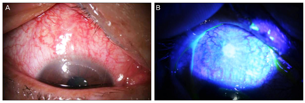
Figure 1 Anterior segment photographs of the right superior bulbar conjunctiva at initial presentation. (A) Slit lamp examination revealed superior conjunctival injection, swelling and corrugation. (B) Fluorescein staining shows epithelial erosion at the superior bulbar conjunctiva.
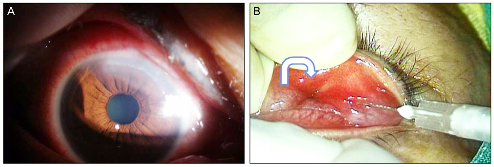
Figure 2 Anterior segment photograph of the cornea near the limbus of the right eye at initial presentation. (A) Slit lamp examination revealed predominant epithelial defect with mild stromal edema. (B) Images were captured from video of subtarsal triamcinolone injection and superior bulbar conjunctival resection of the right eye. Dimpling lesion on the upper eyelid everted with Desmarres lid retractor considered as blepharoplasty scar (curved arrow).
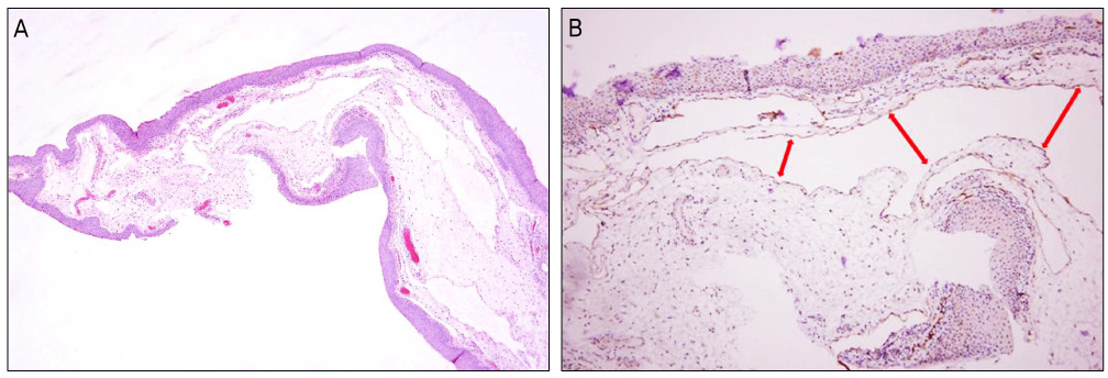
Figure 3 Histopathologic finding of the superior bulbar conjunctiva. (A) Hematoxylin and eosin stain, ×40. (B) Immunohistochemical studies with D2-40 and showing positive staining (brown color) for lymphatic endothelium, ×100. The double headed arrows indicate dilated lymphatic vessels.
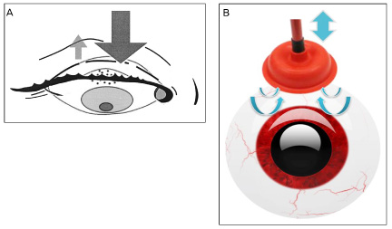
Figure 4 Respective influences of eyelid movements during blink. directions of eyelid movement are indicated by the arrows. (A) Protraction of the palpebral conjunctiva by hypertrophic lesion during the blink has enough force to squeeze the globe (and bulbar conjunctiva) backwards, so has ample power to propel bulbar conjunctiva towards the cornea. (B) Diagrammatic representation of forces producing suction (like using plunger). Dimpling lesion of the palpebral conjunctiva during eye opening has force to create a reciprocating motion along an axis, which then builds pressure in a cylinder. The pressure in the dimpling site actuates the suction force o bulbar conjunctiva.
Cited by 1 articles
-
A Case of Isolated Conjunctival Lymphangioma Mimicking a Recurrent Conjunctival Cyst
Jehwi Jeon, Chan Ho Cho, Sang-Bumm Lee
J Korean Ophthalmol Soc. 2018;59(7):676-679. doi: 10.3341/jkos.2018.59.7.676.
Reference
-
1. Kim HB, Kim EW, Lee JB. Superior limbic keratoconjunctivitis. J Korean Ophthalmol Soc. 1981. 22:395–398.2. Nelson JD. Superior limbic keratoconjunctivitis. Eye. 1989. 3:180–189.3. Cher I. Blink-related microtrauma: when the ocular surface harms itself. Clin Experiment Ophthalmol. 2003. 31:183–190.4. Morax S, Touitou V. Complications of blepharoplasty. Orbit. 2006. 25:303–318.5. Kim JT, Kim JH, Kim JC. Visualization of subconjunctival lymphatics and its significance. J Korean Ophthalmol Soc. 2008. 49:1215–1219.6. Singh D. Conjunctival lymphatic system. J Cataract Refract Surg. 2003. 29:632–633.7. Spector JA, Zide BM. Carbon dioxide laser ablation for treatment of lymphangioma of the conjunctiva. Plast Reconstr Surg. 2006. 117:609–612.8. Wiegand S, Eivazi B, Barth PJ, et al. Pathogenesis of lymphangiomas. Virchows Arch. 2008. 453:1–8.9. Theodore FH, Ferry AP. Superior limbic keratoconjunctivitis: clinical and pathological correlations. Arch Ophthalmol. 1970. 84:481–484.10. Cher I. Superior limbic keratoconjunctivitis: multifactorial mechanical pathogenesis. Clin Experiment Ophthalmol. 2000. 28:181–184.11. Sheu MC, Schoenfield L, Jeng BH. Development of superior limbic keratoconjunctivitis after upper eyelid blepharoplasty surgery:support for the mechanical theory of its pathogenesis. Cornea. 2007. 26:490–492.12. Ohashi Y, Watanabe H, Kinoshita S, et al. Vitamin A eyedrops for superior limbic keratoconjunctivitis. Am J Ophthalmol. 1988. 105:523–527.13. Shen YC, Wang CY, Tsai HY, Lee YF. Supratarsal triamcinolone injection in the treatment of superior limbic keratoconjunctivitis. Cornea. 2007. 26:423–426.14. Duke-Elder S. System of Ophthalmology. 1961. Vol. II & VIII. St. Louis: CV Mosby;541–550. 1965;39-46.15. Joo JH, Ko MK, Park MH. Ultrastructural and immunofluorescent features of lymphatic disorders in conjunctiva. J Korean Ophthalmol Soc. 1987. 28:545–550.
- Full Text Links
-
- Actions
-
Cited
- CITED
-
- Close
- Share
-
- Similar articles
-
- A Case of Conjunctival Lithiasis with Clinical Manifestations of Superior Limbic Keratoconjunctivitis
- Superior Limbic Keratoconjunctivits
- The Change of Eyebrow Position After Upper Lid Blepharoplasty in Patients With Dermatochalasis
- A Case of Primary Lid Tuberculosis after Upper Lid Blepharoplasty
- Two Cases of Superior Limbic Keratoconjunctivitis Treated with Bevacizumab and Triamcinolone Injection
