J Adv Prosthodont.
2015 Apr;7(2):129-137. 10.4047/jap.2015.7.2.129.
Relationship between squamous cell carcinoma of the tongue and the position of dental prosthesis
- Affiliations
-
- 1Department of Oral and Maxillofacial Surgery, Dental Reaserch Institute, Seoul National University, Seoul, Republic of Korea. myoungh@snu.ac.kr myungkim@snu.ac.kr
- KMID: 2118243
- DOI: http://doi.org/10.4047/jap.2015.7.2.129
Abstract
- PURPOSE
Squamous cell carcinoma (SCC) of the tongue has a relatively high incidence of all oral cancers. Some studies have reported a relationship between intraoral dental prosthesis and SCC of the tongue; however, this relationship remains controversial. The purpose of this study was to investigate the relationship between SCC of the tongue and the positional aspects of dental prosthesis using a retrospective analysis.
MATERIALS AND METHODS
A total of 439 patients with SCC of the tongue were diagnosed and treated in the Department of Oral and Maxillofacial Surgery, Seoul National University Dental Hospital. Patients were treated over a 12.5-year period ranging from January 1, 2001 to June 30, 2013. Statistical analysis was performed to examine potential differences between the groups.
RESULTS
The number of patients with a crown and/or a bridge (134, 63.5%) was significantly different than the number of patients without a prosthesis (77, 36.5%). Even after accounting for different types of prostheses such as crowns, bridges, and dentures, no significant differences were observed between the position of the prosthesis and the location of the SCC of the tongue, with significance defined as a P-value less than .05 by the Pearson-Chi square test.
CONCLUSION
Patients with crowns and/or bridges exhibited more frequent SCC of the tongue compared with patients without these prosthesis. These data support the hypothesis that mechanical trauma and galvanic phenomena play a role in the etiology of SCC of the tongue.
MeSH Terms
Figure
-
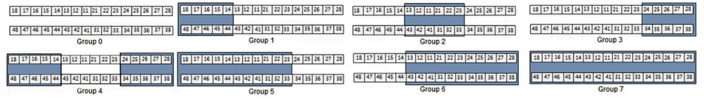
Fig. 1 Group classification according to the tooth positions of crowns and bridges used in this study. Tooth numbers are in accordance with the FDI (Federation Dentaire Internationale) system. Group 0: no crown or bridge, Group 1: right molar area (from the right first premolar to the right third molar), Group 2: anterior teeth area (from the right canine to the left canine), Group 3: left molar area (from the left first premolar to the left third molar), Group 4: bilateral molar area, Group 5: right molar and anterior teeth areas, Group 6: anterior teeth and left molar areas, Group 7: right molar, anterior teeth, and left molar areas.
Reference
-
1. Warnakulasuriya S. Global epidemiology of oral and oropharyngeal cancer. Oral Oncol. 2009; 45:309–316.2. Kolokythas A. Long-term surgical complications in the oral cancer patient: a comprehensive review. Part I. J Oral Maxillofac Res. 2010; 1:e1.3. Byers RM. Squamous cell carcinoma of the oral tongue in patients less than thirty years of age. Am J Surg. 1975; 130:475–478.4. Jones JB, Lampe HB, Cheung HW. Carcinoma of the tongue in young patients. J Otolaryngol. 1989; 18:105–108.5. Llewellyn CD, Johnson NW, Warnakulasuriya KA. Risk factors for squamous cell carcinoma of the oral cavity in young people-a comprehensive literature review. Oral Oncol. 2001; 37:401–418.6. Mashberg A, Boffetta P, Winkelman R, Garfinkel L. Tobacco smoking, alcohol drinking, and cancer of the oral cavity and oropharynx among U.S. veterans. Cancer. 1993; 72:1369–1375.7. Boffetta P, Mashberg A, Winkelmann R, Garfinkel L. Carcinogenic effect of tobacco smoking and alcohol drinking on anatomic sites of the oral cavity and oropharynx. Int J Cancer. 1992; 52:530–533.8. Schwartz SM, Daling JR, Doody DR, Wipf GC, Carter JJ, Madeleine MM, Mao EJ, Fitzgibbons ED, Huang S, Beckmann AM, McDougall JK, Galloway DA. Oral cancer risk in relation to sexual history and evidence of human papillomavirus infection. J Natl Cancer Inst. 1998; 90:1626–1636.9. Laronde DM, Hislop TG, Elwood JM, Rosin MP. Oral cancer: just the facts. J Can Dent Assoc. 2008; 74:269–272.10. Preston-Martin S, Henderson BE, Pike MC. Descriptive epidemiology of cancers of the upper respiratory tract in Los Angeles. Cancer. 1982; 49:2201–2207.11. Cox B, Taylor K, Treasure E. Trends in oral cancer by subsite in New Zealand. Eur J Cancer B Oral Oncol. 1995; 31B:113–117.12. Macfarlane GJ, Sharp L, Porter S, Franceschi S. Trends in survival from cancers of the oral cavity and pharynx in Scotland: a clue as to why the disease is becoming more common? Br J Cancer. 1996; 73:805–808.13. Moore SR, Johnson NW, Pierce AM, Wilson DF. The epidemiology of tongue cancer: a review of global incidence. Oral Dis. 2000; 6:75–84.14. Miyamoto S, Sakuraba M, Nagamatsu S, Kayano S, Kamizono K, Hayashi R. Risk factors for gastric-tube dependence following tongue reconstruction. Ann Surg Oncol. 2012; 19:2320–2326.15. Markopoulos AK. Current aspects on oral squamous cell carcinoma. Open Dent J. 2012; 6:126–130.16. Goldstein DP, Irish JC. Head and neck squamous cell carcinoma in the young patient. Curr Opin Otolaryngol Head Neck Surg. 2005; 13:207–211.17. Jainkittivong A, Aneksuk V, Langlais RP. Oral mucosal conditions in elderly dental patients. Oral Dis. 2002; 8:218–223.18. Lockhart PB, Norris CM Jr, Pulliam C. Dental factors in the genesis of squamous cell carcinoma of the oral cavity. Oral Oncol. 1998; 34:133–139.19. Kinnebrew M, Gettleman L, Carr RF, Beazley R. Squamous cell carcinoma of the tongue in a young woman. Report of a case with etiologic considerations. Oral Surg Oral Med Oral Pathol. 1984; 58:696–698.20. Gorsky M, Silverman S Jr. Denture wearing and oral cancer. J Prosthet Dent. 1984; 52:164–166.21. Jainkittivong A, Aneksuk V, Langlais RP. Oral mucosal lesions in denture wearers. Gerodontology. 2010; 27:26–32.22. Alburqueque R, López-López J, Marí-Roig A, Jané-Salas E, Chimenos-Küstner E, Santos JR. Relationship between squamous cell carcinoma of the anterior two thirds of the tongue and removable denture use: a pioneer study in a Portuguese population. Braz Dent J. 2011; 22:410–414.23. Sharp GS. Treatment for low tolerance to dentures: supplemental report. J Prosthet Dent. 1967; 17:222–226.24. Saito T, Sugiura C, Hirai A, Notani K, Totsuka Y, Shindoh M, Fukuda H. Development of squamous cell carcinoma from pre-existent oral leukoplakia: with respect to treatment modality. Int J Oral Maxillofac Surg. 2001; 30:49–53.25. Korraah A, Odenthal M, Kopp M, Vigneswaran N, Sacks PG, Dienes HP, Stützer H, Niedermeier W. Induction of apoptosis and up-regulation of cellular proliferation in oral leukoplakia cell lines inside electric field. Oral Surg Oral Med Oral Pathol Oral Radiol. 2012; 113:644–654.26. Reinhard MC, Solomon HA. Electrical currents from dental metals as an etiologic factor in oral cancer. Am J Cancer. 1934; 22:606–610.27. Bascom PW. Oral cancer and prosthodontics. J Prosthet Dent. 1968; 19:164–173.
- Full Text Links
-
- Actions
-
Cited
- CITED
-
- Close
- Share
-
- Similar articles
-
- A Case of Basaloid Squamous Cell Carcinoma Occurring in the Mobile Tongue
- The Dental Factors in Oral Squamous Cell Carcinoma
- Glossopharyngeal Neuralgia Secondary to Tongue Squamous Cell Carcinoma
- Expression of E-Cadherin and Matrix Metalloproteinase-2 in Squamous Cell Carcinoma of the Oral Tongue
- The Role of 18F-FDG PET/CT for Evaluation of Cervical Metastatic Lymph Nodes in a Patient with Metallic Artifacts from Dental Prosthesis: a Case Report
