Push-out bond strengths of fiber-reinforced composite posts with various resin cements according to the root level
- Affiliations
-
- 1Department of Conservative Dentistry, Dental Science Research Institute, School of Dentistry, Chonnam National University, Gwangju, Republic of Korea.
- 2Department of Conservative Dentistry, College of Dentistry, Wonkwang University, Iksan, Republic of Korea.
- 3Department of Conservative Dentistry, Wonju College of Medicine, Yonsei University, Wonju, Republic of Korea.
- 4Department of Conservative Dentistry, School of Dentistry, Chonbuk National University, Jeonju, Republic of Korea.
- 5Department of Dental Biomaterials and Institute of Biomaterials & Implant, College of Dentistry, Wonkwang University, Iksan, Republic of Korea. baejimy@wku.ac.kr
- KMID: 2118167
- DOI: http://doi.org/10.4047/jap.2013.5.3.278
Abstract
- PURPOSE
The aim of this study was to determine whether the push-out bond strengths between the radicular dentin and fiber reinforced-composite (FRC) posts with various resin cements decreased or not, according to the coronal, middle or apical level of the root.
MATERIALS AND METHODS
FRC posts were cemented with one of five resin cement groups (RelyX Unicem: Uni, Contax with activator & LuxaCore-Dual: LuA, Contax & LuxaCore-Dual: Lu, Panavia F 2.0: PA, Super-Bond C&B: SB) into extracted human mandibular premolars. The roots were sliced into discs at the coronal, middle and apical levels. Push-out bond strength tests were performed with a universal testing machine at a crosshead speed of 0.5 mm/min, and the failure aspect was analyzed.
RESULTS
There were no significant differences (P>.05) in the bond strengths of the different resin cements at the coronal level, but there were significant differences in the bond strengths at the middle and apical levels (P<.05). Only the Uni and LuA cements did not show any significant decrease in their bond strengths at all the root levels (P>.05); all other groups had a significant decrease in bond strength at the middle or apical level (P<.05). The failure aspect was dominantly cohesive at the coronal level of all resin cements (P<.05), whereas it was dominantly adhesive at the apical level.
CONCLUSION
All resin cement groups showed decreases in bond strengths at the middle or apical level except LuA and Uni.
MeSH Terms
Figure
-
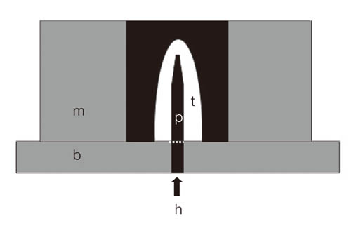
Fig. 1 The cemented post in a tooth embedded in acrylic resin. The end of post was fixed into precisely fitted hole using an aluminum base (p: post, t: tooth, m: aluminum mold, b: aluminum base, h: fitting hole).
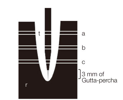
Fig. 2 The FRC post luted with resin cement inside the root canal was embedded in acrylic resin and sectioned at 1 mm thickness producing 3 specimens from the coronal (a), middle (b), and apical areas (c), respectively. At least 3 mm of gutta-percha was left to maintain the apical seal (t: tooth, r: acrylic resin).
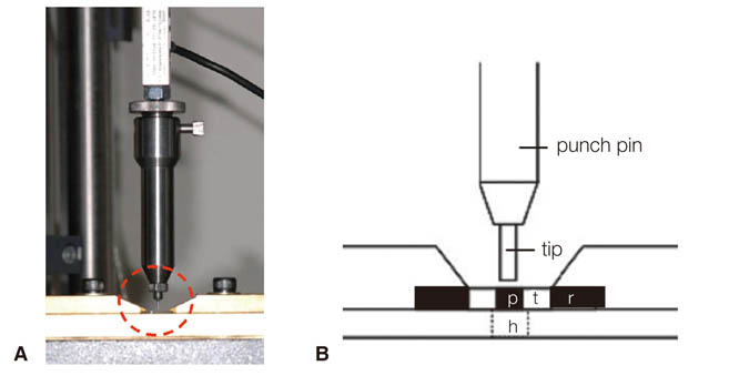
Fig. 3 Assembly of the push-out bond strength test. (A) photograph of the assembly installed on a universal testing machine, (B) a magnified schematic diagram of the circled area of (A) (p: post, t: tooth, r: acrylic resin, h: hole for the post being pushed-out on loading).
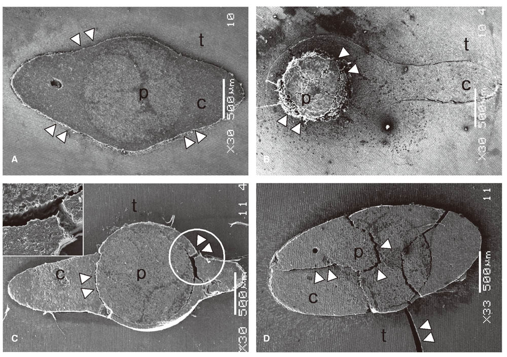
Fig. 4 Representative SEM failure images shown at ×30 maginfication: (A) adhesive failure between dentin and cement at the apical level of PA, (B) adhesive failure between post and cement at the middle level of SB, (C) adhesive failure between post and cement and cohesive failure within cement at the middle level of Uni; the circled area is shown at ×300 in the upper left corner, (D) cohesive failure within post, cement and tooth at the coronal level of LuA (Δ: failure, p: post, c: cement, t: tooth).
Reference
-
1. Schwartz RS, Robbins JW. Post placement and restoration of endodontically treated teeth: a literature review. J Endod. 2004; 30:289–301.2. Sorensen JA, Engelman MJ. Ferrule design and fracture resistance of endodontically treated teeth. J Prosthet Dent. 1990; 63:529–536.3. Akkayan B, Gülmez T. Resistance to fracture of endodontically treated teeth restored with different post systems. J Prosthet Dent. 2002; 87:431–437.4. Bae JM, Kim KN, Hattori M, Hasegawa K, Yoshinari M, Kawada E, Oda Y. Fatigue strengths of particulate filler composites reinforced with fibers. Dent Mater J. 2004; 23:166–174.5. Jung SH, Min KS, Chang HS, Park SD, Kwon SN, Bae JM. Microleakage and fracture patterns of teeth restored with different posts under dynamic loading. J Prosthet Dent. 2007; 98:270–276.6. Mezzomo E, Massa F, Libera SD. Fracture resistance of teeth restored with two different post-and-core designs cemented with two different cements: an in vitro study. Part I. Quintessence Int. 2003; 34:301–306.7. Drummond JL, Toepke TR, King TJ. Thermal and cyclic loading of endodontic posts. Eur J Oral Sci. 1999; 107:220–224.8. Mannocci F, Ferrari M, Watson TF. Intermittent loading of teeth restored using quartz fiber, carbon-quartz fiber, and zirconium dioxide ceramic root canal posts. J Adhes Dent. 1999; 1:153–158.9. Viguie G, Malquarti G, Vincent B, Bourgeois D. Epoxy/carbon composite resins in dentistry: mechanical properties related to fiber reinforcements. J Prosthet Dent. 1994; 72:245–249.10. Vallittu PK. Prosthodontic treatment with a glass fiber-reinforced resin-bonded fixed partial denture: A clinical report. J Prosthet Dent. 1999; 82:132–135.11. Duarte S Jr, Botta AC, Meire M, Sadan A. Microtensile bond strengths and scanning electron microscopic evaluation of self-adhesive and self-etch resin cements to intact and etched enamel. J Prosthet Dent. 2008; 100:203–210.12. Krämer N, Lohbauer U, Frankenberger R. Adhesive luting of indirect restorations. Am J Dent. 2000; 13:60D–76D.13. Özcan M, Mese A. Adhesion of conventional and simplified resin-based luting cements to superficial and deep dentin. Clin Oral Investig. 2012; 16:1081–1088.14. Li N, Nikaido T, Takagaki T, Sadr A, Makishi P, Chen J, Tagami J. The role of functional monomers in bonding to enamel: acid-base resistant zone and bonding performance. J Dent. 2010; 38:722–730.15. Ramos MB, Pegoraro TA, Pegoraro LF, Carvalho RM. Effects of curing protocol and storage time on the microhardness of resin cements used to lute fiber-reinforced resin posts. J Appl Oral Sci. 2012; 20:556–562.16. Sigemori RM, Reis AF, Giannini M, Paulillo LA. Curing depth of a resin-modified glass ionomer and two resin-based luting agents. Oper Dent. 2005; 30:185–189.17. Ritter AV, Ghaname E, Pimenta LA. Dentin and enamel bond strengths of dual-cure composite luting agents used with dual-cure dental adhesives. J Dent. 2009; 37:59–64.18. Bell AM, Lassila LV, Kangasniemi I, Vallittu PK. Bonding of fibre-reinforced composite post to root canal dentin. J Dent. 2005; 33:533–539.19. Benetti AR, Asmussen E, Peutzfeldt A. Influence of curing rate of resin composite on the bond strength to dentin. Oper Dent. 2007; 32:144–148.20. Bitter K, Meyer-Lueckel H, Priehn K, Kanjuparambil JP, Neumann K, Kielbassa AM. Effects of luting agent and thermocycling on bond strengths to root canal dentine. Int Endod J. 2006; 39:809–818.21. Bitter K, Priehn K, Martus P, Kielbassa AM. In vitro evaluation of push-out bond strengths of various luting agents to tooth-colored posts. J Prosthet Dent. 2006; 95:302–310.22. D'Arcangelo C, Cinelli M, De Angelis F, D'Amario M. The effect of resin cement film thickness on the pullout strength of a fiber-reinforced post system. J Prosthet Dent. 2007; 98:193–198.23. Mosharraf R, Baghaei Yazdi N. Comparative evaluation of effects of different surface treatment methods on bond strength between fiber post and composite core. J Adv Prosthodont. 2012; 4:103–108.24. Khamverdi Z, Talebian R. Effect of ascorbic acid, ethanol and acetone on adhesion between the treated fiber posts and composite resin cores. J Adv Prosthodont. 2012; 4:187–191.25. International Standards Organization. ISO/TS 11405: 2003(E) Dental materials - Testing of adhesion to tooth structure. Geneva: ISO;2003.26. Vallittu PK. Flexural properties of acrylic resin polymers reinforced with unidirectional and woven glass fibers. J Prosthet Dent. 1999; 81:318–326.27. Carvalho CA, Breschi L, Navarro MF, Atta MT, Ferrari M. Push-out bond strength and SEM evaluation of a new bonding approach into the root canal. J Appl Oral Sci. 2012; 20:613–619.28. Gaston BA, West LA, Liewehr FR, Fernandes C, Pashley DH. Evaluation of regional bond strength of resin cement to endodontic surfaces. J Endod. 2001; 27:321–324.29. Foxton RM, Nakajima M, Tagami J, Miura H. Adhesion to root canal dentine using one and two-step adhesives with dual-cure composite core materials. J Oral Rehabil. 2005; 32:97–104.30. Pereira PC, Melo RM, Chaves C, Galhano GA, Bottino MA, Balducci I. The adhesive system and root canal region do not influence the degree of conversion of dual resin cement. J Appl Oral Sci. 2010; 18:477–481.31. Teixeira CS, Silva-Sousa YT, Sousa-Neto MD. Bond strength of fiber posts to weakened roots after resin restoration with different light-curing times. J Endod. 2009; 35:1034–1039.32. Mallmann A, Jacques LB, Valandro LF, Mathias P, Muench A. Microtensile bond strength of light- and self-cured adhesive systems to intraradicular dentin using a translucent fiber post. Oper Dent. 2005; 30:500–506.33. Bouillaguet S, Troesch S, Wataha JC, Krejci I, Meyer JM, Pashley DH. Microtensile bond strength between adhesive cements and root canal dentin. Dent Mater. 2003; 19:199–205.34. Perdigão J, Gomes G, Lee IK. The effect of silane on the bond strengths of fiber posts. Dent Mater. 2006; 22:752–758.35. Acquaviva PA, Cerutti F, Adami G, Gagliani M, Ferrari M, Gherlone E, Cerutti A. Degree of conversion of three composite materials employed in the adhesive cementation of indirect restorations: a micro-Raman analysis. J Dent. 2009; 37:610–615.36. Feilzer AJ, de Gee AJ, Davidson CL. Setting stresses in composites for two different curing modes. Dent Mater. 1993; 9:2–5.37. Kato H, Matsumura H, Atsuta M. Effect of etching and sandblasting on bond strength to sintered porcelain of unfilled resin. J Oral Rehabil. 2000; 27:103–110.38. Radovic I, Monticelli F, Goracci C, Vulicevic ZR, Ferrari M. Self-adhesive resin cements: a literature review. J Adhes Dent. 2008; 10:251–258.39. Barkmeier WW, Cooley RL. Laboratory evaluation of adhesive systems. Oper Dent. 1992; Suppl 5. 50–61.40. Staninec M, Kawakami M. Adhesion and microleakage tests of a new dentin bonding system. Dent Mater. 1993; 9:204–208.41. Watanabe I, Nakabayashi N, Pashley DH. Bonding to ground dentin by a phenyl-P self-etching primer. J Dent Res. 1994; 73:1212–1220.42. Gomes AL, Castillo-Oyagüe R, Lynch CD, Montero J, Albaladejo A. Influence of sandblasting granulometry and resin cement composition on microtensile bond strength to zirconia ceramic for dental prosthetic frameworks. J Dent. 2013; 41:31–41.
- Full Text Links
-
- Actions
-
Cited
- CITED
-
- Close
- Share
-
- Similar articles
-
- Evaluation of the resin cement thicknesses and push-out bond strengths of circular and oval fiber posts in oval-shapes canals
- The effect of different adhesive system applications on push-out bond strengths of glass fiber posts
- Effect of different adhesive systems and post surface treatments on the push-out bond strengths of fiber-reinforced post
- Effects of post surface conditioning before silanization on bond strength between fiber post and resin cement
- Effect of antioxidants on push-out bond strength of hydrogen peroxide treated glass fiber posts bonded with two types of resin cement
