Infiltrating Epidural Angiolipoma Involving Lumbar Spine
- Affiliations
-
- 1Department of Orthopedic Surgery, Barun Orthopedics Hospital, Busan, Korea.
- 2Department of Orthopedic Surgery, Busan Catholic Maryknoll Medical Center, Busan, Korea. dr.leehyeongseok@gmail.com
- KMID: 2106751
- DOI: http://doi.org/10.4055/jkoa.2015.50.2.148
Abstract
- We report on an unusual case with infiltrating extradural spinal angiolipoma. Most spinal angiolipomas involve the thoracic spine and infiltrating ones are also located mainly at the thoracic levels rather than lumbar lesion. In particular, there are few cases of lumbar extradural infiltrating type spinal angiolipoma. One case is that of a 52-year-old female with infiltrating extradural spinal angiolipoma involving lumbar 4 (L4) vertebra, who underwent a L4-5 laminectomy and surgical removal of the tumor. We achieved satisfactory results with surgical treatment of the patient. Spinal angiolipoma has a benign course with a good postoperative outcome.
Keyword
Figure
-
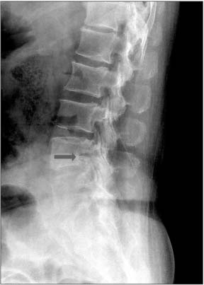
Figure 1 Preoperative plain radiograph shows a radiolucent lesion in lumbar 4 vertebral body (arrow).

Figure 2 Computed tomography (CT) scan and magnetic resonance imaging show a well-defined fatty mass at lumbar 4 (L4) vertebral body with bony erosion. (A) CT scan showing bony erosion of the right posterior L4 vertebral body. (B) T-1 weighted sagittal and axial images showing a hyperintense fatty mass in the right anterior epidural space. (C) T-2 weighted sagittal and axial images showing hypointense fatty mass in the right anterior epidural space.

Figure 3 T-1 weighted sagittal and axial images with gadolinium showing a hyperintense fatty mass in the right anterior epidural space.
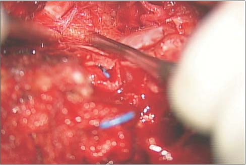
Figure 4 Gross photograph shows a yellow-gray and soft-to-firm tumor in the epidural area-intraoperatively.

Figure 5 Microscopic photograph shows mature adipose tissue interspersed with well vascularized areas, indicative of angiolipoma. Hypovascularized area suggestive of a conventional lipoma (H&E, ×200).
Reference
-
1. Ehni G, Love JG. Intraspinal Lipomas Report of cases; Review of the literature, and clinical and pathologic study. Arch Neurol Psychiatry. 1945; 53:1–28.2. Preul MC, Leblanc R, Tampieri D, Robitaille Y, Pokrupa R. Spinal angiolipomas. Report of three cases. J Neurosurg. 1993; 78:280–286.3. Lin JJ, Lin F. Two entities in angiolipoma. A study of 459 cases of lipoma with review of literature on infiltrating angiolipoma. Cancer. 1974; 34:720–727.4. Fourney DR, Tong KA, Macaulay RJ, Griebel RW. Spinal angiolipoma. Can J Neurol Sci. 2001; 28:82–88.
Article5. Leu NH, Chen CY, Shy CG, et al. MR imaging of an infiltrating spinal epidural angiolipoma. AJNR Am J Neuroradiol. 2003; 24:1008–1011.6. Shin BJ, Kim KJ, Suh YS, Kim DW. Epidural angiolipoma: a case report. J Korean Soc Spine Surg. 1997; 4:165–169.7. Park JH, Jeon SR, Rhim SC, Roh SW. Lumbar spinal extradural angiolipoma: case report and review of the literature. J Korean Neurosurg Soc. 2008; 44:265–267.8. Rocchi G, Caroli E, Frati A, Cimatti M, Savlati M. Lumbar spinal angiolipomas: report of two cases and review of the literature. Spinal Cord. 2004; 42:313–316.
Article9. Turgut M. Spinal angiolipomas: report of a case and review of the cases published since the discovery of the tumour in 1890. Br J Neurosurg. 1999; 13:30–40.
Article10. Fourney DR, Tong KA, Macaulay RJ, Griebel RW. Spinal angiolipoma. Can J Neurol Sci. 2001; 28:82–88.
Article
