Airway Compression as a Result of Extensive Prevertebral Hematoma Following Extension Injury of Lower Cervical Spine without Fracture/Dislocation
- Affiliations
-
- 1Department of Orthopedic Surgery, Chonbuk National University Medical School, Chonbuk National University Hospital, Jeonju, Korea. osdr2815@naver.com
- 2Research Institute of Clinical Medicine, Chonbuk National University Hospital, Jeonju, Korea.
- KMID: 2106653
- DOI: http://doi.org/10.4055/jkoa.2012.47.3.227
Abstract
- A 77-year-old man presented with severe dyspnea, neck pain, tingling sensation in both hands, and weakness after an acute prevertebral soft tissue hematoma due to distractive-extension injury. Magnetic resonance images demonstrated an extensive hematoma accumulation, anterior longitudinal ligament and longus colli muscle injuries. We report here a case of dyspnea due to an extensive prevertebral hematoma by soft tissue injury without cervical vertebral fracture and/or dislocation and a review the relevant literature.
MeSH Terms
Figure
-
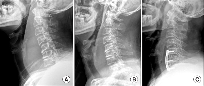
Figure 1 (A) Initial C-spine lateral radiograph shows extensive prevertebral soft tissue shadow (retropharyngeal space at C3: 29.3 mm, retrotracheal space at C6: 53.9 mm) without fracture/dislocation from C1 to thoracic area. The retropharyngeal space lies between the pharynx the pharynx and the cervical spine. (B) Post-trauma 1 week C-spine lateral radiograph shows the decrease of prevertebral soft tissue shadow compare to initial C-spine lateral radiograph. (C) Immediate post-operative C-spine lateral radiograph showed marked decrease of prevertebral soft tissue shadow and anterior cervical discectomy and fusion with plate and cage construct at C5-7.
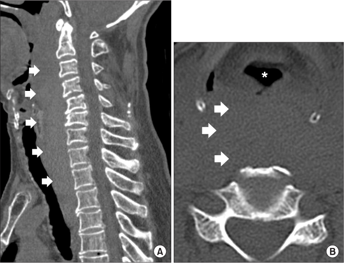
Figure 2 Sagittal (A) and axial (B) computed tomography images show the compression and deviation of trachea (asterix) by extensive prevertebral soft tissue shadow (arrows) and no bony abnormality.
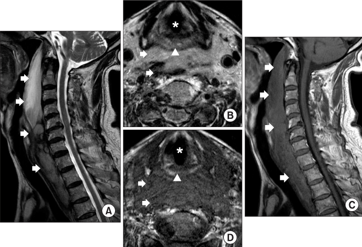
Figure 3 Sagittal (A, C) and axial (B, D) T1, T2-weighted magnetic resonance images show the deviation and compression of trachea (asterix) and esophagus (arrow head) by extensive hematoma in the prevertebral space (arrows) from C1 to T4, the tear of anterior longitudinal ligament and longus coli muscle, disc herniation C5-6, C6-7.
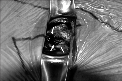
Figure 4 Operative photo shows the extensive hematoma and longus coli muscle tear (white arrows).
Reference
-
1. Emery SE, Smith MD, Bohlman HH. Upper-airway obstruction after multilevel cervical corpectomy for myelopathy. J Bone Joint Surg Am. 1991. 73:544–551.
Article2. Howcroft AJ, Jenkins DH. Potentially fatal asphyxia following a minor injury of the cervical spine. J Bone Joint Surg Br. 1977. 59:93–94.
Article3. Kuhn JE, Graziano GP. Airway compromise as a result of retropharyngeal hematoma following cervical spine injury. J Spinal Disord. 1991. 4:264–269.
Article4. O'Donnell JJ, Birkinshaw R, Harte B. Mechanical airway obstruction secondary to retropharyngeal haematoma. Eur J Emerg Med. 1997. 4:166–168.5. Silberstein M, Tress BM, Hennessy O. Prevertebral swelling in cervical spine injury: identification of ligament injury with magnetic resonance imaging. Clin Radiol. 1992. 46:318–323.
Article6. Fauci AS, Longo DL. Fauci AS, Braunwald E, Kasper DL, editors. Critical care medicine. Harrison's principles of internal medicine. 2008. 17th Edition. New York: The McGraw-Hill Companies;1681–1685.
- Full Text Links
-
- Actions
-
Cited
- CITED
-
- Close
- Share
-
- Similar articles
-
- A Case of Delayed Airway Obstruction Associated with Prevertebral Hematoma and Cervical Vertebra Fracture
- Airway Obstruction Caused by Soft Tissue Edema during an Anterior Cervical Approach: A Case Report
- Cervical Prevertebral Hematoma - a Rare Complication of Acupuncture Therapy: A Case Report
- Management of Cervical Spine Injuries without Fracture or Dislocation
- Traumatic Retropharyngeal Hematoma following Cervical Vascular Injury: A Case Report
