J Korean Orthop Assoc.
2008 Apr;43(2):207-212. 10.4055/jkoa.2008.43.2.207.
The Change of the Posterior Tibial Slope after Cruciate Retaining Total Knee Arthroplasty
- Affiliations
-
- 1Department of Orthopedic Surgery, School of Medicine, Kyung Hee University, Seoul, Korea. bdkyung@khmc.or.kr
- KMID: 2106441
- DOI: http://doi.org/10.4055/jkoa.2008.43.2.207
Abstract
-
PURPOSE: To analyze the pre- and postoperative posterior tibial slope angle (PSA) of performing cruciate-retaining total knee arthroplasty (TKA) and to identify the ideal value of the PSA in relation to the clinical results.
MATERIALS AND METHODS
From June 1999 to May 2005, 202 TKA with a NexGen(R) cruciate-retaining knee were performed in 160 patients. The mean follow-up period was 39.8 months. The pre- and postoperative PSA referenced by the proximal tibial medullary canal (PSA-A) and the proximal tibial anterior cortex (PSA-B) were measured by two independent observers. The knee and function scoring system of the American Knee Society and the range of motion of the knee at the last follow-up were evaluated as the clinical results.
RESULTS
The mean PSA-A was 11.4+/-4.8degrees preoperatively and 6.0+/-2.8degrees postoperatively, and the mean PSA-B was 13.6+/-4.9degrees preoperatively and 8.1+/-2.9degrees postoperatively. The difference between the pre- and postoperative PSA increased as the preoperative PSA-A changed from 6.0degrees and the PSA-B changed from 8.1degrees; these findings showed statistical significance based on a simple linear regression (PSA-A: r= 0.837, p=0.000; PSA-B: r=0.834, p=0.000). The knee and function score of American Knee Society improved respectively from 62.9 and 55.8, preoperatively, to 97.4 and 89.7 respectively, at the last follow-up. The range of motion of the knee joint was 128.0degrees preoperatively and 129.7degrees at the last follow-up.
CONCLUSION
In cruciate retaining total knee arthroplasty, PSA-A is mostly distributed within 3.2-8.8degrees, and a reasonable range of PSA-A is 6.0+/-2.8degrees.
Figure
-
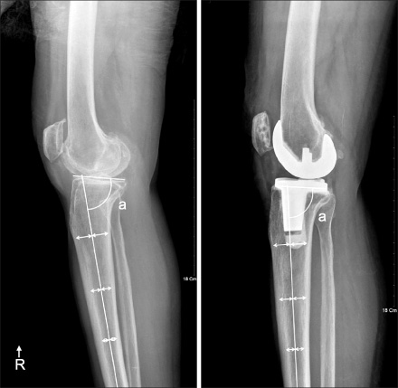
Fig. 1 The measure method of the proximal tibial medullary canal-referenced posterior slope (posterior slope: 90-a). The reference line is the best fit line to 3 points, each of which is the center of the medullary canal just below the tibial tuberosity and at 10 cm and 15 cm distal to the tibial medial plateau.

Fig. 2 The measure method of the proximal tibial anterior cortex-referenced posterior slope (posterior slope: 90-a). The reference line is parallel to the anterior cortex below the tibial tuberosity.
Reference
-
1. Bai B, Baez J, Testa N, Kummer FJ. Effect of posterior cut angle on tibial component loading. J Arthroplasty. 2000. 15:916–920.
Article2. Bellemans J, Robijns F, Duerinckx J, Banks S, Vandenneucker H. The influence of tibial slope on maximal flexion after total knee arthroplasty. Knee Surg Sports Traumatol Arthrosc. 2005. 13:193–196.
Article3. Brazier J, Migaud H, Gougeon F, Cotten A, Fontaine C, Duquennoy A. Evaluation of methods for radiographic measurement of the tibial slope. A study of 83 healthy knees. Rev Chir Orthop Reparatrice Appar Mot. 1996. 82:195–200.4. Buechel FF, Pappas MJ. New Jersey low contact stress knee replacement system. Ten-year evaluation of meniscal bearings. Orthop Clin North Am. 1989. 20:147–177.5. Catani F, Fantozzi S, Ensini A, Leardini A, Moschella D, Giannini S. Influence of tibial component posterior slope on in vivo knee kinematics in fixed-bearing total knee arthroplasty. J Orthop Res. 2006. 24:581–587.
Article6. Chiu KY, Zhang SD, Zhang GH. Posterior slope of tibial plateau in Chinese. J Arthroplasty. 2000. 15:224–227.
Article7. Choi CH, Kim JH, Chung HK, Choi YH. Measurement of posterior slope angle of the proximal tibia by MRI and X-ray. J Korean Orthop Assoc. 2001. 36:569–573.
Article8. Dejour H, Bonnin M. Tibial translation after anterior cruciate ligament rupture. Two radiological tests compared. J Bone Joint Surg Br. 1994. 76:745–749.
Article9. Dixon MC, Brown RR, Parsch D, Scott RD. Modular fixed-bearing total knee arthroplasty with retention of the posterior cruciate ligament. A study of patients followed for a minimum of fifteen years. J Bone Joint Surg Am. 2005. 87:598–603.10. Dorr LD, Boiardo RA. Technical considerations in total knee arthroplasty. Clin Orthop Relat Res. 1986. 5–11.
Article11. Ewald FC, Jacobs MA, Miegel RE, Walker PS, Poss R, Sledge CB. Kinematic total knee replacement. J Bone Joint Surg Am. 1984. 66:1032–1040.
Article12. Giffin JR, Vogrin TM, Zantop T, Woo SL, Harner CD. Effects of increasing tibial slope on the biomechanics of the knee. Am J Sports Med. 2004. 32:376–382.
Article13. Hofmann AA, Bachus KN, Wyatt RW. Effect of the tibial cut on subsidence following total knee arthroplasty. Clin Orthop Relat Res. 1991. 63–69.
Article14. Insall JN, Dorr LD, Scott RD, Scott WN. Rationale of the Knee Society clinical rating system. Clin Orthop Relat Res. 1989. 13–14.
Article15. Jenny JY, Rapp E, Kehr P. Proximal tibial meniscal slope: a comparison with the bone slope. Rev Chir Orthop Reparatrice Appar Mot. 1997. 84:435–438.16. Jiang CC, Yip KM, Liu TK. Posterior slope angle of the medial tibial plateau. J Formos Med Assoc. 1994. 93:509–512.17. Jojima H, Whiteside LA, Ogata K. Effect of tibial slope or posterior cruciate ligament release on knee kinematics. Clin Orthop Relat Res. 2004. 194–198.
Article18. Kuwano T, Urabe K, Miura H, et al. Importance of the lateral anatomic tibial slope as a guide to the tibial cut in total knee arthroplasty in Japanese patients. J Orthop Sci. 2005. 10:42–47.
Article19. Laskin RS, Riegèr MA. The surgical technique for performing a total knee replacement arthroplasty. Orthop Clin North Am. 1989. 20:31–48.20. Massin P, Gournay A. Optimization of the posterior condylar offset, tibial slope, and condylar roll-back in total knee arthroplasty. J Arthroplasty. 2006. 21:889–896.
Article21. Matsuda S, Miura H, Nagamine R, et al. Posterior tibial slope in the normal and varus knee. Am J Knee Surg. 1999. 12:165–168.22. Piazza SJ, Delp SL, Stulberg SD, Stern SH. Posterior tilting of the tibial component decreases femoral rollback in posterior-substituting knee replacement: a computer simulation study. J Orthop Res. 1998. 16:264–270.
Article23. Robertson DD, Yuan J, Bigliani LU, Flatow EL, Yamaguchi K. Three-dimensional analysis of the proximal part of the humerus: relevance to arthroplasty. J Bone Joint Surg Am. 2000. 82:1594–1602.
Article24. Sokal RR, Rohlf FJ. Biometry. 1995. 3rd ed. New York: William H. Freeman;460–461.25. Walker PS, Garg A. Range of motion in total knee arthroplasty. A computer analysis. Clin Orthop Relat Res. 1991. 262:227–235.26. Wasielewski RC, Galante JO, Leighty RM, Natarajan RN, Rosenberg AG. Wear patterns on retrieved polyethylene tibial inserts and their relationship to technical considerations during total knee arthroplasty. Clin Orthop Relat Res. 1994. 299:31–43.
Article27. Whiteside LA, Amador DD. The effect of posterior tibial slope on knee stability after Ortholoc total knee arthroplasty. J Arthroplasty. 1988. 3 Suppl. S51–S57.
Article
- Full Text Links
-
- Actions
-
Cited
- CITED
-
- Close
- Share
-
- Similar articles
-
- Relationship Between ground Surface and Tibial Posterior Slope at Erect Posture in Total Knee Arthroplasty
- Effect of Posterior Femoral Condylar Offset and Posterior Tibial Slope on Maximal Flexion Angle of the Knee in Posterior Cruciate Ligament Sacrificing Total Knee Arthroplasty
- Influence of Posterior Tibial Slope on Stability after Total Knee Arthroplasty
- Influence of the Posterior Slope of the Tibial Component on the Maximal Flexion after Total Knee Arthroplasty
- Changes in Femoral Posterior Condylar Offset, Tibial Posterior Slope Angle, and Joint Line Height after Cruciate-Retaining Total Knee Arthroplasty
