Lateral Closing Wedge Supracondylar Osteotomy of the Humerus in Children with Cubitus Varus Deformity
- Affiliations
-
- 1Department of Orthopedic Surgery, SungKyunKwan University School of Medicine, Seoul, Korea. jss3505@skku.edu
- KMID: 2106411
- DOI: http://doi.org/10.4055/jkoa.2008.43.1.17
Abstract
-
PURPOSE: To evaluate the effectiveness, cosmetic and functional improvement of a supracondylar lateral closing wedge osteotomy of the humerus as a treatment for cubitus varus deformity in children.
MATERIALS AND METHODS
Forty-eight children with cubitus varus underwent a lateral closing wedge osteotomy, and were followed up for at least 1 year.
RESULTS
There were no complications such as a loss of correction, infection, or neurapraxia. The immediate postoperative lateral condylar prominence and secondary lazy S deformity was in proportion to the preoperative severity of the cubitus varus. However, it was lower at the last follow-up, and was related to the extent of preoperative cubitus varus, length of follow-up and age.
CONCLUSION
A supracondylar lateral closing wedge osteotomy of humerus is an easy and effective surgical treatment for a posttraumatic cubitus varus of children. In addition, it shows good cosmetic results with good remodeling of the lateral condylar prominence of children.
Figure
-

Fig. 1 (A) Lateral closing wedge osteotomy was performed. (B) Lazy 'S' deformity was observed on the radiograph of two months after the operation.

Fig. 2 (A) Humero-ulnar angle was measured on the anteroposterior radiograph of the elbow. (B) Shaft-condylar angle was measured on the lateral radiograph of the elbow.
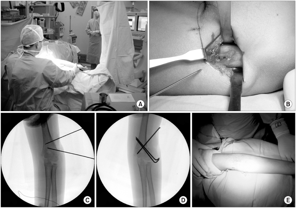
Fig. 3 (A) The angle of cubitus varus deformity was measured intraoperatively with C-arm fluoroscopy. (B) The operation was performed through a lateral approach. (C) We used two preset Kirschner's wires. (D) Osteotomized site was fixed with Steinmann pins. (E) Cubitus varus was corrected after surgery.

Fig. 4 (A) Lateral prominence index (LPI) is BC/AB. (B) Lateral prominence amount (LPA) is BC/AC × 100 (%).

Fig. 5 (A) A six-year-old boy had the varus humeroulnar angle of 33° on the preoperative radiograph. (B) On the immediate postoperative radiograph, LPI was 3.23 and LPA was 45.3 percent. (C) On the radiograph after a 4-year-follow-up, the LPI was 1.01, and the LPA was zero percent.

Fig. 6 Lateral prominence amount decreased with the increasing follow-up duration.

Fig. 7 The change in the amount of lateral prominence diminished with increasing age when the operation had been performed.
Reference
-
1. Barrett IR, Bellemore MC, Kwon YM. Cosmetic results of supracondylar osteotomy for correction of cubitus varus. J Pediatr Orthop. 1998. 18:445–447.
Article2. Bellemore MC, Barrett IR, Middleton RW, Scougall JS, Whiteway DW. Supracondylar osteotomy of the humerus for correction of cubitus varus. J Bone Joint Surg Br. 1984. 66:566–572.
Article3. Danielsson LG, Hussein S, el-Haddad I, Gupta RP. Staple fixation of osteotomy for cubitus varus. A simple technique used in II children. Acta Orthop Scand. 1991. 62:55–57.4. Devnani AS. Lateral closing wedge supracondylar osteotomy of humerus for post-traumatic cubitus varus in children. Injury. 1997. 28:643–647.
Article5. Graham B, Tredwell SJ, Beauchamp RD, Bell HM. Supracondylar osteotomy of the humerus for correction of cubitus varus. J Pediatr Orthop. 1990. 10:228–231.
Article6. Griffin PP. Supracondylar fractures of the humerus. Pediatr Clin North Am. 1975. 22:477–486.
Article7. Høyer A. Treatment of supracondylar fracture of the humerus by skeletal traction in an abduction splint. J Bone Joint Surg Am. 1952. 24:623–637.8. Hui JP, Torode IP, Chatterjee A. Medial approach for corrective osteotomy of cubitus varus: a cosmetic incision. J Pediatr Orthop. 2004. 24:477–481.9. Ippolito E, Moneta MR, D'Arrigo C. Post-traumatic cubitus varus. Long-term follow-up of corrective supracondylar humeral osteotomy in children. J Bone Joint Surg Am. 1990. 72:757–765.
Article10. Karatosun V, Alekberov C, Alici E, Ardic CO, Aksu G. Treatment of cubitus varus using the Ilizarov technique of distraction osteogenesis. J Bone Joint Surg Br. 2000. 82:1030–1033.
Article11. King D, Secor C. Bow elbow(cubitus varus). J Bone Joint Surg Am. 1951. 33:572–576.12. Kumar K, Sharma VK, Sharma R, Maffulli N. Correction of cubitus varus by French or dome osteotomy: A comparative study. J Trauma. 2000. 49:717–721.
Article13. LaBelle H, Bunnell WP, Duhaime M, Poitras B. Cubitus varus deformity following supracondylar osteotomy of the humerus in children. J Pediatr Orthop. 1982. 2:539–540.14. Levin MJ, Horn BD, Pizzultillo PD. Treatment of posttraumatic cubitus varus in the pediatric population with humeral osteotomy and external fixation. J Pediatr Orthop. 1996. 16:597–601.15. McCoy GF, Piggot J. Supracondylar osteotomy for cubitus varus. J Bone Joint Surg Br. 1988. 70:283–286.16. Oppenheim WL, Clader TJ, Smith C, Bayer M. Supracondylar humeral osteotomy for traumatic childhood cubitus varus deformity. Clin Orthop Relat Res. 1984. 188:34–39.
Article17. Pankaj A, Dua A, Malhotra R, Bhan S. Dome osteotomy for posttraumatic cubitus varus: A surgical technique to avoid lateral condylar prominence. J Pediatr Orthop. 2006. 26:61–66.18. Tachdjian MR. Smith AB, editor. Osteotomy for distal humerus for correction of cubitus varus. Pediatric orthopedics. 1972. Philadelphia: Saunders WB;1588–1591.19. Tien YC, Chih HW, Lin GT, Lin SY. Dome corrective osteotomy for cubitus varus deformity. Clin Orthop Relat Res. 2000. 380:158–166.
Article20. Usui M, Ishii S, Miyano S, Narita H, Kura H. Three-dimensional corrective osteotomy for treatment of cubitus varus after supracondylar fracture of the humerus in children. J Shoulder Elbow Surg. 1995. 4:17–22.
Article21. Voss FR, Kasser JR, Trepman E, Simmons E, Hall JE. Uniplanar supracondylar humeral osteotomy with preset Kirschner wires for posttraumatic cubitus varus. J Pediatr Orthop. 1994. 14:471–478.
Article22. Williams PL, Warwick R. Gray's anatomy. 1980. 36th ed. Philadelphia: WB Saunders Co;365.23. Wong HK, Lee EH, Balasubramaniam P. The lateral condylar prominence: a complication of supracondylar osteotomy for cubitus varus. J Bone Joint Surg Br. 1990. 72:859–861.
Article
- Full Text Links
-
- Actions
-
Cited
- CITED
-
- Close
- Share
-
- Similar articles
-
- Supracondylar Osteotomy for Cubitus Vnrus Deformity by Using Plate in Adults
- Supracondylar Quadrilateral Displacement Osteotomy for Cubitus Varus Deformity: New Operative Technique
- Supracondylar Osteotomy in Cubitus Varus and Cubitus Valgus
- Supracondlylar Osteotomy for Cubitus Varus
- The Supracondylar Osteotomy for the Angular Dformity followed by a Fracture Around the Elbow
