T1-weighted FLAIR MR Imaging for the Evaluation of Enhancing Brain Tumors: Comparison with Spin Echo Imaging
- Affiliations
-
- 1Department of Radiology, Gyeongsang National University School of Medicine, Jinju, Korea. choids@gnu.ac.kr
- 2Gyeongsang Institute of Health Science, Gyeongsang National University School of Medicine, Jinju, Korea.
- KMID: 2099891
- DOI: http://doi.org/10.13104/jksmrm.2014.18.2.151
Abstract
- PURPOSE
Spin-echo (SE) technique is most commonly used pulse sequence for T1-weighted MR imaging. T1-weighted fluid-attenuated inversion recovery (T1FLAIR) is a relatively new pulse sequence and it provides higher tissue contrast between the gray matter (GM) and white matter (WM) of the brain than T1-weighted SE (T1SE) sequence. However, there has been controversy for the evaluation of enhancing brain tumors with T1FLAIR compared to T1SE. The purpose of this study was to compare T1FLAIR and T1SE sequences for the evaluation of enhancing intracranial tumors.
MATERIALS AND METHODS
Fifty-two patients with enhancing brain tumors were evaluated with contrast-enhanced (CE) T1SE and T1FLAIR imaging. Eight quantitative criteria were calculated: lesion-to-WM contrast ratio (CR) and contrast-to-noise ratio (CNR), lesion-to-GM CR and CNR, lesion-to-CSF CR and CNR, and WM-to-GM CR and CNR. For qualitative evaluation, two radiologists assessed lesion conspicuity on CE T1SE and T1FLAIR sequences with three-scale: 1, T1SE superior; 2, sequence equal; T1FLAIR superior.
RESULTS
Seventy-nine tumors (31 primaries, 48 metastases) were assessed. For quantitative measurement, the T1FLAIR lesion-to-GM, lesion-to-CSF, WM-to-GM CR and CNR values were comparable and statistically superior to those of the T1SE images (p < 0.001 in all). However, lesion-to-WM CR and CNR were similar on both two sequences without statistically significant difference (p = 0.661, 0.662, respectively). For qualitative evaluation, both radiologists assessed that T1FLAIR images were superior to T1SE images for the evaluation of lesion conspicuity.
CONCLUSION
For the evaluation of enhancing intracranial tumors, T1FLAIR sequence was superior or comparable to T1SE sequence.
Keyword
Figure
-

Fig. 1 A 44-year-old man with a meningioma in the anterior cranial fossa. Contrast-enhanced T1FLAIR (a) and T1SE (b) images show an enhancing tumor in the anterior cranial fossa. The gray-white matter contrast and lesion conspicuity are better on FLAIR image than on SE image.

Fig. 2 A 47-year-old woman with a metastatic tumor from small cell lung cancer. Contrast-enhanced T1FLAIR (a) and T1SE (b) images show a metastatic enhancing lesion in the left frontal white matter. The gray-white matter contrast and lesion conspicuity are better on FLAIR image than on SE image. The superior lesion conspicuity on FLAIR image might be caused by the suppression of water signal intensity in the surrounding brain edema.
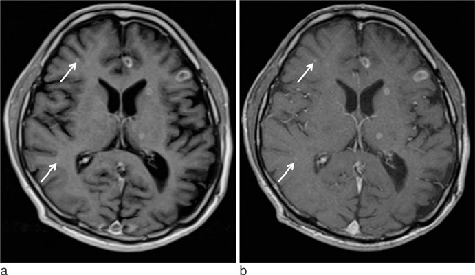
Fig. 3 A 52-year-old man with multiple metastatic tumors from small cell lung cancer. Contrast-enhanced T1FLAIR (a) and T1SE (b) images show multiple enhancing lesions in both frontal and right temporal lobes and left basal ganglia and thalamus. The conspicuity of the left frontal cortical lesions is superior on T1FLAIR compared to that on T1SE. However, the right frontal and temporal lobes lesions (arrows) and the left basal ganglia and thalamic lesions are more clearly demonstrated on T1SE image than on T1FLAIR image.
Cited by 1 articles
-
Advances in magnetic resonance technique for tumor imaging
Dong Woo Park
J Korean Med Assoc. 2015;58(6):516-522. doi: 10.5124/jkma.2015.58.6.516.
Reference
-
1. Qian YF, Yu CL, Zhang C, Yu YQ. MR T1-weighted inversion recovery imaging in detecting brain metastases: could it replace T1-weighted spin-echo imaging? AJNR Am J Neuroradiol. 2008; 29:701–704.2. Melhem ER, Israel DA, Eustace S, Jara H. MR of the spine with a fast T1-weighted fluid-attenuated inversion recovery sequence. AJNR Am J Neuroradiol. 1997; 18:447–454.3. Al-Saeed O, Ismail M, Athyal RP, Rudwan M, Khafajee S. T1-weighted fluid-attenuated inversion recovery and T1-weighted fast spin-echo contrast-enhanced imaging: a comparison in 20 patients with brain lesions. J Med Imaging Radiat Oncol. 2009; 53:366–372.4. Rydberg JN, Hammond CA, Huston J 3rd, Jack CR Jr, Grimm RC, Riederer SJ. T1-weighted MR imaging of the brain using a fast inversion recovery pulse sequence. J Magn Reson Imaging. 1996; 6:356–362.5. Lee JK, Choi HY, Lee SW, Baek SY, Kim HY. Usefulness of T1-weighted image with fast inversion recovery technique in intracranial lesions: comparison with T1-weighted spin echo image. Clin Imaging. 2000; 24:263–269.6. Melhem ER, Bert RJ, Walker RE. Usefulness of optimized gadolinium-enhanced fast fluid-attenuated inversion recovery MR imaging in revealing lesions of the brain. AJR Am J Roentgenol. 1998; 171:803–807.7. Fischbach F, Bruhn H, Pech M, et al. Efficacy of contrast medium use for neuroimaging at 3.0 T: utility of IR-FSE compared to other T1-weighted pulse sequences. J Comput Assist Tomogr. 2005; 29:499–450.
- Full Text Links
-
- Actions
-
Cited
- CITED
-
- Close
- Share
-
- Similar articles
-
- T1-, T2-weighted, and FLAIR Imaging: Clinical Application
- Fast FLAIR MR Images of Intracranial Hemorrhage
- Usefulness of Fluid Attenuated Inve rsion Re c overy(FLAIR) Image
- Fast FLAIR MR Imaging Finidngs of Cerebral Infarction: Comparison with T2-Weighted Spin Echo Imaging
- Hyperacute Intracerebral Hemorrhage: Comparison of EPI and Other MR Sequences
