Intradiploic Encephalocele of the Left Parietal Bone: A Case Report
- Affiliations
-
- 1Department of Neurosurgery, Myongji St. Mary's Hospital, Seoul, Korea. nshs@catholic.ac.kr
- 2Department of Radiology, Myongji St. Mary's Hospital, Seoul, Korea.
- KMID: 2098023
- DOI: http://doi.org/10.3348/jksr.2015.72.6.423
Abstract
- Encephaloceles are generally regarded as midline abnormalities. A 50-year-old man presented with a parietal intradiploic encephalocele manifesting as intermittent headache for the past 6 months. Computed tomography (CT) showed bone destruction associated with a left parietal lesion. Magnetic resonance imaging (MRI) demonstrated brain herniation within the intradiploic space. Cerebral angiographic imaging showed a normal cerebral vessel pattern within the herniated brain lesion. In this case, surgical treatment may not be necessary in the absence of concurrent symptoms and neurologic deficit. We report the CT, MRI, and angiographic findings of an extremely rare case of parietal intradiploic encephalocele in adulthood.
MeSH Terms
Figure
-
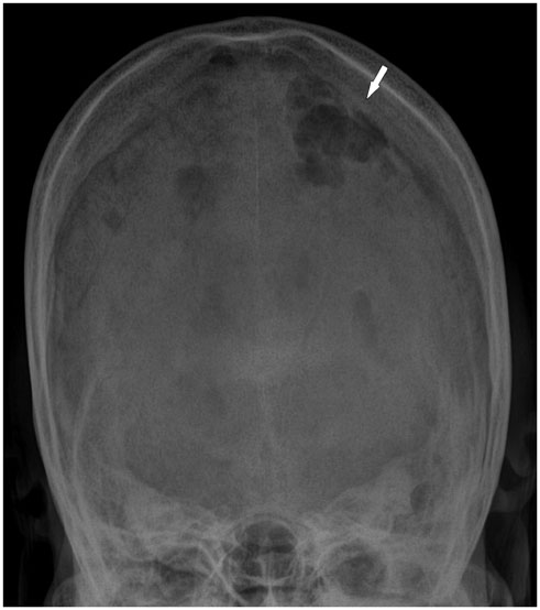
Fig. 1 Skull film: town view shows the left parietal defect (white arrow).
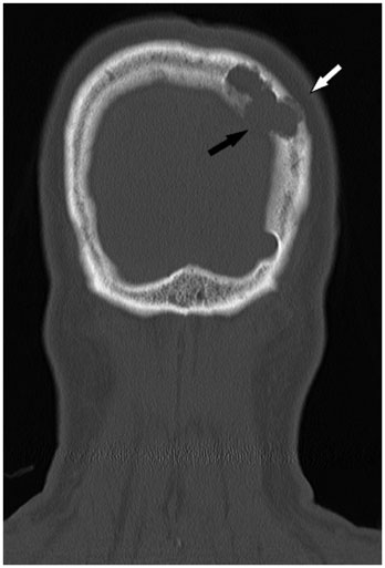
Fig. 2 Coronal image of bone window CT shows a defect of the inner table, widened diploic space (black arrow). There is marked thinning and erosion of the outer table of the left parietal parasagittal bone (white arrow).
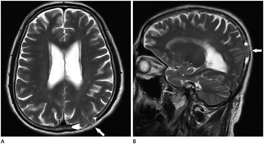
Fig. 3 MRI shows a lesion in the parietal parasagittal intradiploic space continuous with the left parietal lobe appearing isointense with the normal brain on axial T2-weighted (A) image. Sagittal T2-weighted MRI shows a defect in the left parietal bone, which contains cerebrospinal fluid and herniated cerebral tissue (white arrow) (B).

Fig. 4 Left internal carotid artery angiogram with arterial (A) and venous (B) phase films show normal cerebral vessel pattern for herniated brain in left parietal bony defect area (black arrowheads).
Reference
-
1. Kosnik EJ, Meagher JN, Quenemoen LR. Parietal intradiploic encephalocele. Case report. J Neurosurg. 1976; 44:617–661.2. Mealey J Jr, Dzenitis AJ, Hockey AA. The prognosis of encephaloceles. J Neurosurg. 1970; 32:209–218.3. Tsuboi Y, Hayashi N, Noguchi K, Kurimoto M, Nagai S, Endo S. Parietal intradiploic encephalocele--case report. Neurol Med Chir (Tokyo). 2007; 47:240–224.4. Sadler TW. Langman's medical embryology. 10th ed. Baltimore: Lippincott Williams & Wilkins;2006. p. 126–143. p. 286–317.5. Peters J, Raab P, Marquardt G, Zanella FE. Intradiploic meningoencephalocele. Eur Radiol. 2002; 12:Suppl 3. S25–S27.6. Dobrin N, Bălinişteanu M, Costăchescu B, Tudorache C, Chiriac A, Poeată I. Acquired parietal intradiploic encephalocele. Case report and review of the literature. Rom S Neurosurg. 2011; 18.7. Loumiotis I, Jones L, Diehn F, Lanzino G. Symptomatic left intradiploic encephalocele. Neurology. 2010; 75:1027.8. Kumar R, Chandra SP, Sharma BS. Giant intradiploic pseudomeningocele of occipital bone. J Neurosurg Pediatr. 2012; 9:82–85.
