Fulminant Superior Ophthalmic Vein and Cavernous Sinus Thrombophlebitis with Intracranial Extensions: Case Reports
- Affiliations
-
- 1Department of Radiology, Soonchunhyang University Bucheon Hospital, Soonchunhyang University College of Medicine, Bucheon, Korea. hshong@schmc.ac.kr
- 2Department of Infectious Diseases, Soonchunhyang University Bucheon Hospital, Soonchunhyang University College of Medicine, Bucheon, Korea.
- KMID: 2098022
- DOI: http://doi.org/10.3348/jksr.2015.72.6.418
Abstract
- Cavernous sinus thrombophlebitis (CST) is a rare and life-threatening disease without prompt diagnosis and treatment. Two cases of fulminant superior ophthalmic vein (SOV) and CST caused by maxillary periodontitis and sphenoid sinusitis are described. A 65-year-old woman presented with right proptosis, headache, and fever. A 74-year-old woman presented with left periorbital swelling. In both patients, MRI with gadolinium showed expansion of the bilateral cavernous sinus and diffuse dilatation of the SOV with non-enhancement of central thrombus, which indicated CST. The condition was complicated by brain abscess, meningitis, and ischemic stroke. These conditions were improved by antibiotic treatment, but one patient underwent exenteration of the orbit due to orbital rupture during hospitalization.
MeSH Terms
-
Aged
Brain Abscess
Cavernous Sinus
Cavernous Sinus Thrombosis*
Diagnosis
Dilatation
Exophthalmos
Female
Fever
Gadolinium
Headache
Hospitalization
Humans
Magnetic Resonance Imaging
Meningitis
Orbit
Paranasal Sinus Diseases
Periodontitis
Rupture
Sphenoid Sinus
Sphenoid Sinusitis
Stroke
Thrombophlebitis
Thrombosis
Veins*
Gadolinium
Figure
-

Fig. 1 A 65-year-old female presented with 2-week history of right-sided proptosis, headache, and fever. A. Axial contrast-enhanced three-dimensional fast spoiled gradient-echo (3D FSPGR) image reveals multifocal, irregular filling defects within an expanded, enhanced cavernous sinus. B. Axial contrast-enhanced 3D FSPGR image shows linear filling defect in dilated left superior ophthalmic vein (SOV). C. The thrombosed left SOV exhibits a high signal intensity on diffusion-weighted imaging. D. Axial contrast-enhanced T1-weighted spin echo images (repetition time/echo time, 600/9.3 msec) shows brain abscesses on the left anterior temporal convexity.
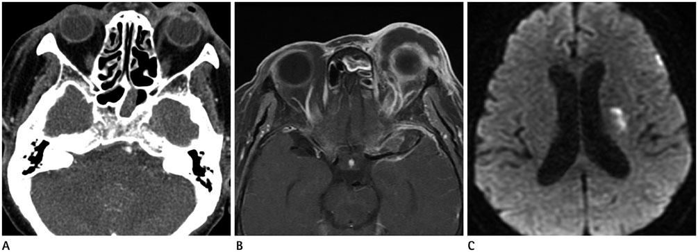
Fig. 2 A 74-year-old female presented with periorbital swelling and purulent discharge from left eye. A. Axial contrast-enhanced computed tomographic scan reveals left-sided proptosis, a gas-containing abscess in the left preseptal region. The left sphenoid sinus exhibits mucosal thickening. B. Axial contrast-enhanced T1-weighted fat-suppressed spin-echo image (repetition time/echo time, 816.6/9 msec) reveals a dilated, non-enhanced, left superior ophthalmic vein and rim-enhanced abscesses in the preseptal region and intraconal space. Thick enhancement of the dura along the left frontotemporal cerebral convexity is seen. C. Follow-up diffusion-weighted imaging after orbital exenteration reveals ischemic infarctions in the left basal ganglia and periventricular white matter.
Reference
-
1. Bhatia K, Jones NS. Septic cavernous sinus thrombosis secondary to sinusitis: are anticoagulants indicated? A review of the literature. J Laryngol Otol. 2002; 116:667–676.2. Osborn AG. Osborn's brain: Imaging, Pathology, and Anatomy. Philadelphia: Lippincott Williams & Wilkins;2012. p. 217–218.3. DiNubile MJ. Septic thrombosis of the cavernous sinuses. Arch Neurol. 1988; 45:567–572.4. Southwick FS, Richardson EP Jr, Swartz MN. Septic thrombosis of the dural venous sinuses. Medicine (Baltimore). 1986; 65:82–106.5. Dolan RW, Chowdhury K. Diagnosis and treatment of intracranial complications of paranasal sinus infections. J Oral Maxillofac Surg. 1995; 53:1080–1087.6. Gallagher RM, Gross CW, Phillips CD. Suppurative intracranial complications of sinusitis. Laryngoscope. 1998; 108(11 Pt 1):1635–1642.7. Lee JH, Lee HK, Park JK, Choi CG, Suh DC. Cavernous sinus syndrome: clinical features and differential diagnosis with MR imaging. AJR Am J Roentgenol. 2003; 181:583–590.8. Razek AA, Castillo M. Imaging lesions of the cavernous sinus. AJNR Am J Neuroradiol. 2009; 30:444–452.9. Schuknecht B, Simmen D, Yüksel C, Valavanis A. Tributary venosinus occlusion and septic cavernous sinus thrombosis: CT and MR findings. AJNR Am J Neuroradiol. 1998; 19:617–626.10. Mahapatra AK. Brain abscess--an unusual complication of cavernous sinus thrombosis. A case report. Clin Neurol Neurosurg. 1988; 90:241–243.
- Full Text Links
-
- Actions
-
Cited
- CITED
-
- Close
- Share
-
- Similar articles
-
- Coil Embolization Via a Superior Ophthalmic Vein Approach of Carotid Cavernous Sinus Fistula
- Treatment of a Carotid-Cavernous Sinus Fistula via the Superior Ophthalmic Vein Approach: A Case Report
- A Case of Cavernous Sinus Thrombophlebitis Secondary toAcute Isolated Sphenoid Sinusitis
- Transvenous Embolization of Cavernous Sinus Dural Arteriovenous Fistula Using the Direct Superior Ophthalmic Vein Approach: A Case Report
- Intraoperative Embolization of Dural Carotid-Cavernous Fistula: Case Report
