Imaging Findings of Signet Ring Cell Carcinoma of the Ampulla of Vater: A Case Report
- Affiliations
-
- 1Department of Radiology, Eulji Hospital, Eulji University School of Medicine, Seoul, Korea. jyy@eulji.ac.kr
- 2Department of Surgery, Eulji Hospital, Eulji University School of Medicine, Seoul, Korea.
- 3Department of Pathology, Eulji Hospital, Eulji University School of Medicine, Seoul, Korea.
- KMID: 2098016
- DOI: http://doi.org/10.3348/jksr.2015.72.6.381
Abstract
- Most malignant tumors affecting the ampulla of Vater (AoV) are adenocarcinomas, and signet ring cell carcinoma (SRCC) of the AoV is rare. We report a case of SRCC of the AoV in a 41-year-old man who presented with a small tumor in the AoV, which was discovered by multiple imaging modalities. Since most of the previously reported articles have been published in the clinical pathology literature, we mainly described the imaging features.
MeSH Terms
Figure
-
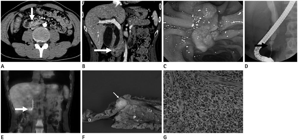
Fig. 1 41-year-old man with signet ring cell carcinoma of the ampulla of Vater. A, B. Axial (A) and coronal (B) reformatted contrast-enhanced CT images in the portal venous phase show a lobulating mass with enhancement (arrows) in the ampulla of Vater. Upstream intrahepatic and extrahepatic bile ducts are dilated. C. Duodenoscopy shows an ulcerofungating mass in the ampulla of Vater, macroscopically. D. Endoscopic retrograde cholangiography shows nodular filling defect (arrow) in the far-distal common bile duct, corresponding to the intraluminal nodular enhancing lesion found on CT. Diffuse dilatation of common bile duct is seen. E. Coronal 18F-FDG PET/CT image shows increased FDG uptake (arrow) in the distal common bile duct without evidence of distant metastases. F. The gross cut surface of surgical specimen shows a whitish periampullary tumor (arrow). The tumor invaded through the duodenal wall but the pancreas is grossly unremarkable. G. Microscopic photomicrograph shows diffusely infiltrative adenocarcinoma. Upper half is composed of usual signet ring cells and lower half is consisted of eosinophilic signet ring cells (hematoxylin & eosin stain, × 400). D = duodenum, P = pancreas, 18F-FDG PET/CT = 18-fluoro-2-deoxy-D-glucose positron emission tomography/computed tomography
Reference
-
1. Kamisawa T, Fukayama M, Koike M, Tabata I, Egawa N, Isawa T, et al. Carcinoma of the ampulla of Vater: expression of cancer-associated antigens inversely correlated with prognosis. Am J Gastroenterol. 1988; 83:1118–1123.2. Albores-Saavedra J, Schwartz AM, Batich K, Henson DE. Cancers of the ampulla of vater: demographics, morphology, and survival based on 5,625 cases from the SEER program. J Surg Oncol. 2009; 100:598–605.3. Talebi A, Mohammadizadeh F, Hani M, Bagheri M, Bagheri A. Signet ring carcinoma of ampulla of vater. Adv Biomed Res. 2014; 3:30.4. Kim S, Lee NK, Lee JW, Kim CW, Lee SH, Kim GH, et al. CT evaluation of the bulging papilla with endoscopic correlation. Radiographics. 2007; 27:1023–1038.5. Mosconi S, Beretta GD, Labianca R, Zampino MG, Gatta G, Heinemann V. Cholangiocarcinoma. Crit Rev Oncol Hematol. 2009; 69:259–270.6. Lee JH, Park MS, Kim KW, Yu JS, Kim MJ, Yang SW, et al. Advanced gastric carcinoma with signet ring cell carcinoma versus non-signet ring cell carcinoma: differentiation with multidetector CT. J Comput Assist Tomogr. 2006; 30:880–884.7. Taghavi S, Jayarajan SN, Davey A, Willis AI. Prognostic significance of signet ring gastric cancer. J Clin Oncol. 2012; 30:3493–3498.8. Lee EY, Kim C, Kim MJ, Park JY, Park SW, Song SY, et al. Signet ring cell carcinoma of the extrahepatic bile duct. Gut Liver. 2010; 4:402–406.9. Gao JM, Tang SS, Fu W, Fan R. Signet-ring cell carcinoma of ampulla of Vater: contrast-enhanced ultrasound findings. World J Gastroenterol. 2009; 15:888–891.10. Chang S, Lim JH, Choi D, Kim SK, Lee WJ. Differentiation of ampullary tumor from benign papillary stricture by thin-section multidetector CT. Abdom Imaging. 2008; 33:457–462.
- Full Text Links
-
- Actions
-
Cited
- CITED
-
- Close
- Share
-
- Similar articles
-
- A Case of Signet-ring Cell Carcinoma of the Ampulla of Vater
- Cytologic Features of Signet Ring Cell Carcinoma of the Uterine Cervix: A Report of Two Cases
- A Composite Tumor of the Ampulla of Vater: Signet-ring Cell and Neuroendocrine Carcinoma: A Case Report
- A Case of a Collision Tumor in the Ampulla of Vater with an Adenocarcinoma and a Large Cell Neuroendocrine Carcinoma
- CT Finding of Signet Ring Cell Carcinoma of the Stomach
