Aneurysm and Infundibular Dilatation at an Unusual Origin of the Ophthalmic Artery
- Affiliations
-
- 1Department of Radiology, Kyung Hee University Hospital, Seoul, Korea. euijkim@hanmail.net
- KMID: 2097999
- DOI: http://doi.org/10.3348/jksr.2014.71.4.164
Abstract
- The ophthalmic artery usually arises from the anteromedial or superomedial aspect of the internal carotid artery. Rarely does it arise from the medial or posteromedial aspect of the internal carotid artery. In this paper, the authors report two cases of aneurysm and infundibular dilatation found at unusual sites of origin in the ophthalmic artery and review the literature about possible etiologies contributing to the anatomic variations.
Figure
-
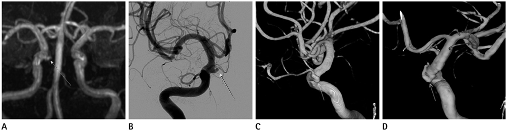
Fig. 1 Unusual origin of ophthalmic artery in a 55-year-old woman. A. Magnetic resonance angiography shows an bulging lesion (arrow) suspected to be an aneurysm at the medial aspect of the ophthalmic segment of the right internal carotid artery (ICA). B. Anteroposterior view of the right carotid angiography shows the bulging lesion (arrow) from the medial aspect of ophthalmic segment of the right ICA. C, D. Lateral view (C) and posterior view (D) of three-dimensional rotational angiography shows a vessel (arrow) arising from the posteromedial aspect of ophthalmic segment of the right ICA and running forward.
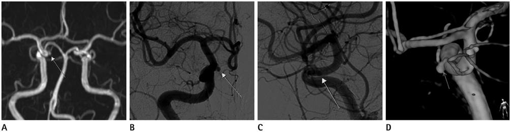
Fig. 2 Unusual origin of ophthalmic artery in a 58-year-old woman. A. Magnetic resonance angiography shows a bulging lesion suspected to be an aneurysm (arrow) at the medial aspect of the ophthalmic segment of the right internal carotid artery (ICA). B, C. Anteroposterior view (B) and lateral view (C) of the right carotid angiography shows an aneurysm (arrow) from the medial aspect of ophthalmic segment of the right ICA. D. Posterior view of three-dimensional rotational angiography shows a small saccular aneurysm (arrow) with an aneurismal neck at carotid-ophthalmic junction.

Fig. 3 Embryology of the ophthalmic artery proposed by Lasjaunias et al. A. The VOA originates from the ACA and the DOA originates from the ICA. B, C. Two anastomoses formed. D. The proximal parts of the VOA and DOA regress. Note.-ACA = anterior cerebral artery, DOA = dorsal ophthalmic artery, ICA = internal carotid artery, OA = ophthalmic artery, VOA = ventral ophthalmic artery

Fig. 4 Embryology of the ophthalmic artery proposed by Padget. A. The DOA appears at the opposite site of the ICA bifurcation. B. The DOA moves proximally. C. The anastomosis between VOA and DOA is formed. D. The dorsal portion of VOA regress. Note.-DOA = dorsal ophthalmic artery, ICA = internal carotid artery, OA = ophthalmic artery, VOA = ventral ophthalmic artery
Reference
-
1. Sade B, Tampieri D, Mohr G. Ophthalmic artery originating from basilar artery: a rare variant. AJNR Am J Neuroradiol. 2004; 25:1730–1731.2. Lasjaunias P, Moret J, Mink J. The anatomy of the inferolateral trunk (ILT) of the internal carotid artery. Neuroradiology. 1977; 13:215–220.3. Padget DH. The development of the cranial arteries in the human embryo. Contrib Embryol. 1948; 32:205–262.4. Hayreh SS, Dass R. The ophthalmic artery: I. origin and intra-cranial and intra-canalicular course. Br J Ophthalmol. 1962; 46:65–98.5. Gibo H, Lenkey C, Rhoton AL Jr. Microsurgical anatomy of the supraclinoid portion of the internal carotid artery. J Neurosurg. 1981; 55:560–574.6. Baltsavias G, Türk Y, Valavanis A. Persistent ventral ophthalmic artery associated with supraclinoid internal carotid artery aneurysm: case report and review of the literature. J Neuroradiol. 2012; 39:186–189.7. Tanaka M. Persistent primitive dorsal ophthalmic artery associated with paraclinoid internal carotid artery aneurysm. JNET. 2009; 3:39–41.8. Locksley HB. Natural history of subarachnoid hemorrhage, intracranial aneurysms and arteriovenous malformations. Based on 6368 cases in the cooperative study. J Neurosurg. 1966; 25:219–239.9. Satoh T, Omi M, Ohsako C, Fujiwara K, Tsuno K, Sasahara W, et al. Differential diagnosis of the infundibular dilation and aneurysm of internal carotid artery: assessment with fusion imaging of 3D MR cisternography/angiography. AJNR Am J Neuroradiol. 2006; 27:306–312.
- Full Text Links
-
- Actions
-
Cited
- CITED
-
- Close
- Share
-
- Similar articles
-
- An Aneurysm Developing on the Infundibulum of Posterior Communicating Artery: Case Report and Literature Review
- Coil Embolization of Traumatic Ophthalmic Artery Aneurysm: Case Report
- A Case of Giant Paraclinoid Aneurysm
- Infundibular Widening of Angiographically Invisible Duplicate Anterior Choroidal Artery Mimicking Typical Anterior Choroidal Artery Aneurysm
- Intracranial Aneurysm Associated with Aplasia of the Internal Cartoid Artery
