Post-Traumatic Contrast Enhancing Brain Lesion
- Affiliations
-
- 1Department of Radiology, Eulji Hospital, Eulji University College of Medicine, Seoul, Korea. khs46359@eulji.ac.kr
- 2Department of Neurosurgery, Eulji Hospital, Eulji University College of Medicine, Seoul, Korea.
- KMID: 2097997
- DOI: http://doi.org/10.3348/jksr.2014.71.4.155
Abstract
- Only a few studies have been reported on the MR contrast enhancement and the apparent diffusion coefficient (ADC) findings of the post-traumatic lesion of the brain. We report a case of the venous ischemia in the left frontal lobe observed in the MRI obtained one day after the incidence of trauma. Considering the presented slight increase in the ADC, the vasogenic edema was thought to be the major mechanism of the venous ischemia and excitotoxic injury. In spite of a slight increase in the ADC, the hyperintensity in the diffusion weighted imaging and contrast-enhanced areas eventually changed into hemorrhagic lesions.
Figure
-
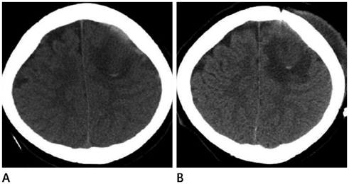
Fig. 1 Initial (A) and 8-days follow-up (B) axial CT images. A. Initial axial CT image shows subdural hematoma and brain swelling in the left frontal lobe with focal hyperattenuated spot, suggesting hemorrhage. B. Eight-days follow-up axial CT image shows progressive white matter edema in the left frontal lobe without gross change that suggests focal hemorrhage in the corticomedullary junction.
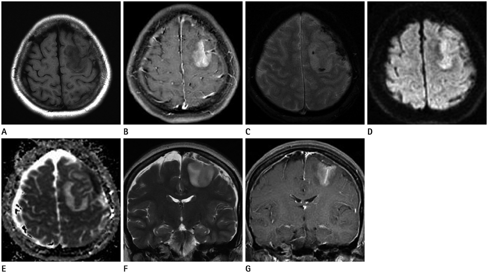
Fig. 2 A 40-year-old woman with acute trauma and seizure. A. Axial T1-weighted MR image shows hypointensity on the left frontal lobe. B. Axial gadolinium-enhanced T1-weighted MR image shows contrast-enhancement of the left frontal lobe. C. Axial gradient-echo MR image shows two hypointensities in the corticomedullary junction of the left frontal lobe, suggesting hemorrhage. D. Axial diffusion-weighted MR image shows cortical hyperintensity in the left frontal lobe. E. Axial apparent diffusion coefficient MR image shows slight cortical hyperintensity in the left frontal lobe with edema of adjacent white matter. F. Coronal T2-weighted MR image shows hyperintensity in the superior and middle frontal gyrus with edema of subcortical white matter. G. Coronal gadolinium-enhanced T1-weighted MR image shows gyral enhancement of the left frontal lobe. Asymmetric high signal intensity in the left side wall and lumen of the anterior superior sagittal sinus is seen.
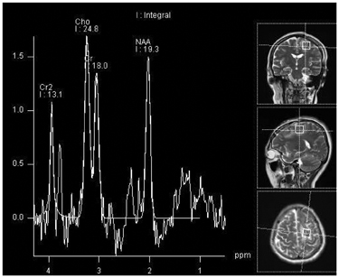
Fig. 3 MR spectroscopy obtained 19 days later. Subtle increase of Choline (Cho)/Creatinine (Cr) ratio is seen to be 1.33.
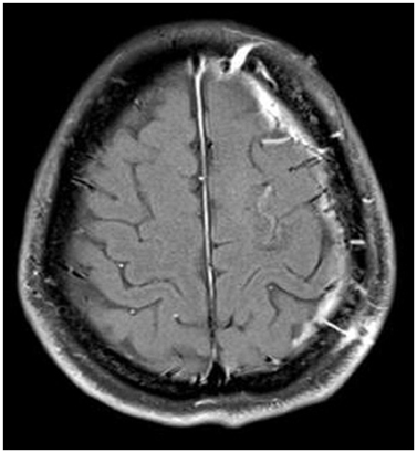
Fig. 4 Follow-up MR image obtained two months later. Axial gadolinium-enhanced T1-weighted image shows a markedly improved left frontal lobe lesion.
Reference
-
1. Matsushige T, Nakaoka M, Kiya K, Takeda T, Kurisu K. Cerebral sinovenous thrombosis after closed head injury. J Trauma. 2009; 66:1599–1604.2. Forbes KP, Pipe JG, Heiserman JE. Evidence for cytotoxic edema in the pathogenesis of cerebral venous infarction. AJNR Am J Neuroradiol. 2001; 22:450–455.3. Osborn AG. Osborn's barin: imaging, pathology and anatomy. Salt Lake City: Amirsys;2013. p. 1–72.4. Stam J. Thrombosis of the cerebral veins and sinuses. N Engl J Med. 2005; 352:1791–1798.5. Saposnik G, Barinagarrementeria F, Brown RD Jr, Bushnell CD, Cucchiara B, Cushman M, et al. Diagnosis and management of cerebral venous thrombosis: a statement for healthcare professionals from the American Heart Association/American Stroke Association. Stroke. 2011; 42:1158–1192.6. Alahmadi H, Vachhrajani S, Cusimano MD. The natural history of brain contusion: an analysis of radiological and clinical progression. J Neurosurg. 2010; 112:1139–1145.7. Armin SS, Colohan AR, Zhang JH. Vasospasm in traumatic brain injury. Acta Neurochir Suppl. 2008; 104:421–425.8. Shahlaie K, Keachie K, Hutchins IM, Rudisill N, Madden LK, Smith KA, et al. Risk factors for posttraumatic vasospasm. J Neurosurg. 2011; 115:602–611.9. Kim JH, Chang KH, Na DG, Song IC, Kwon BJ, Han MH, et al. 3T 1H-MR spectroscopy in grading of cerebral gliomas: comparison of short and intermediate echo time sequences. AJNR Am J Neuroradiol. 2006; 27:1412–1418.10. Leach JL, Fortuna RB, Jones BV, Gaskill-Shipley MF. Imaging of cerebral venous thrombosis: current techniques, spectrum of findings, and diagnostic pitfalls. Radiographics. 2006; 26:Suppl 1. S19–S41. discussion S42-S43.
- Full Text Links
-
- Actions
-
Cited
- CITED
-
- Close
- Share
-
- Similar articles
-
- Delirium After Traumatic Brain Injury: Prediction by Location and Size of Brain Lesion
- Two Cases of Traumatic Pericallosal Artery Aneurysm: A Case Report
- T1-weighted FLAIR MR Imaging for the Evaluation of Enhancing Brain Tumors: Comparison with Spin Echo Imaging
- Comparison of MR Imaging Findings Between Post-operative Change and Residual/Recurrent Tumor in Cerebral Glioma
- Contrast-Enhanced Turbo Spin-Echo(TSE) T1-weighted Imaging: Improved Contrast of Enhancing Lesions
