Chordoma Mimicking Sellar Tumor: A Case Report
- Affiliations
-
- 1Department of Radiology, Gangneung Asan Hospital, College of Medicine, University of Ulsan, Gangneung, Korea. neurorad@lycos.co.kr
- 2Department of Neurology, Gangneung Asan Hospital, College of Medicine, University of Ulsan, Gangneung, Korea.
- 3Department of Pathology, Gangneung Asan Hospital, College of Medicine, University of Ulsan, Gangneung, Korea.
- KMID: 2097975
- DOI: http://doi.org/10.3348/jksr.2012.67.6.413
Abstract
- Chordoma is a rare tumor that accounts for approximately 0.2% all intracranial tumors. We report a pathologically proven chordoma, mimicking a sellar tumor.
MeSH Terms
Figure
-
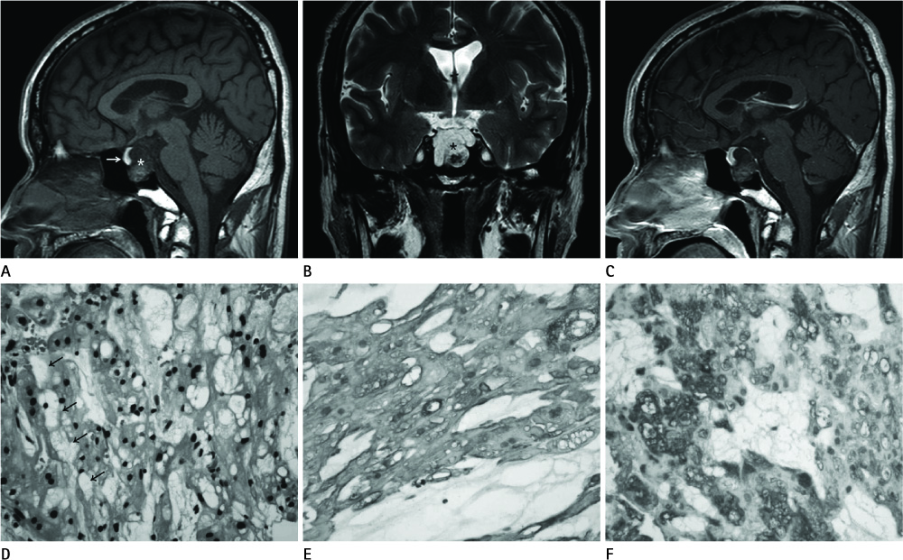
Fig. 1 50-year-old male with chordoma. A. Sagittal T1-weighted (500/10) image shows a tumor (asterisk) in the sellar area, displacing the pituitary gland (arrow) anteriorly. The mass shows heterogeneous low signal intensity. B. Coronal T2-weighted image (3000/80) shows lobulated tumor (asterisk) with heterogeneous high signal intensity. C. Sagittal Gd-DTPA contrast enhanced T1-weighted (500/10) image shows minimal enhancement of a sellar tumor. D. Photomicrograph reveals round vacuolated cells containing intracytoplasmic mucus droplets with lobular arrangement (physaliphorous appearance) (arrows), which is finding of a typical chordoma (H&E stain, × 400). E, F. Immunohistochemical photography reveals tumor cells stained with antibodies to S100 protein and CK, respectively (original magnification, × 200).
Reference
-
1. Choi GM, Han MH, Chang KH, Yu IK, Kim HD, Kim SS, et al. Chordoma versus chondrosarcoma of the central skull base: MR and CT findings. J Korean Radiol Soc. 1998. 38:221–228.2. Lee SK, Han CH, Lee MO, Kim MY, Yi JG, Lee JH, et al. Clival chordoma: CT and MR findings. J Korean Radiol Soc. 1993. 29:687–692.3. Heffelfinger MJ, Dahlin DC, MacCarty CS, Beabout JW. Chordomas and cartilaginous tumors at the skull base. Cancer. 1973. 32:410–420.4. Hirosawa RM, Santos AB, França MM, Fabris VE, Castro AV, Zanini MA, et al. Intrasellar chondroid chordoma: a case report. ISRN Endocrinol. 2011. 2011:259392.5. Rosenberg AE, Brown GA, Bhan AK, Lee JM. Chondroid chordoma--a variant of chordoma. A morphologic and immunohistochemical study. Am J Clin Pathol. 1994. 101:36–41.6. Meyer JE, Oot RF, Lindfors KK. CT appearance of clival chordomas. J Comput Assist Tomogr. 1986. 10:34–38.7. Whelan MA, Reede DL, Meisler W, Bergeron RT. CT of the base of the skull. Radiol Clin North Am. 1984. 22:177–217.8. Sze G, Uichanco LS 3rd, Brant-Zawadzki MN, Davis RL, Gutin PH, Wilson CB, et al. Chordomas: MR imaging. Radiology. 1988. 166(1 Pt 1):187–191.9. Doucet V, Peretti-Viton P, Figarella-Branger D, Manera L, Salamon G. MRI of intracranial chordomas. Extent of tumour and contrast enhancement: criteria for differential diagnosis. Neuroradiology. 1997. 39:571–576.10. Lanzino G, Dumont AS, Lopes MB, Laws ER Jr. Skull base chordomas: overview of disease, management options, and outcome. Neurosurg Focus. 2001. 10:E12.
