Primary Follicular Carcinoma Arising in Ectopic Thyroid Tissue of the Lateral Neck: A Case Report
- Affiliations
-
- 1Department of Radiology, College of Medicine, Hanyang University, Korea. dwpark@hanyang.ac.kr
- 2Department of Pathology, College of Medicine, Hanyang University, Korea.
- KMID: 2097910
- DOI: http://doi.org/10.3348/jksr.2010.63.5.413
Abstract
- Ectopic thyroid tissue in the lateral neck is an uncommon congenital anomaly, and the occurrence of primary follicular carcinoma in this ectopic thyroid tissue is very rare. We report here on such a case of follicular carcinoma arising in ectopic thyroid tissue of the left lateral neck without any evidence of primary carcinoma in the original thyroid gland.
Figure
-

Fig. 1 CT findings on the lateral neck mass in a 39-year-old man. A. The axial pre-enhanced CT image shows a 4.2×3.0×5.4 cm-sized welldefined, isodense mass (arrow) in the left posterior submandibular space. B. The axial postcontrast CT image shows the strong and inhomogenous enhancement of the mass (arrow). The left submandibular gland is displaced anteriorly by the mass.
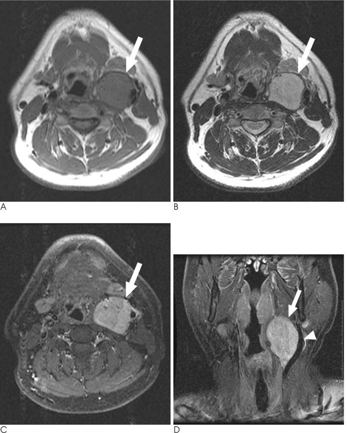
Fig. 2 MRI findings on the lateral neck mass. A, B. The mass (arrow) shows well-defined, homogenous isointensity on the T1-weighted MR image (A) and mild hyperintensity on the T2-weighted MR image (B). C, D. The axial and coronal fat-saturated gadolinium-enhanced T1-weighted MR images show inhomogeneous enhancement of the mass (arrow). The external and internal carotid arteries are displaced laterally due to the mass (arrowhead in D).
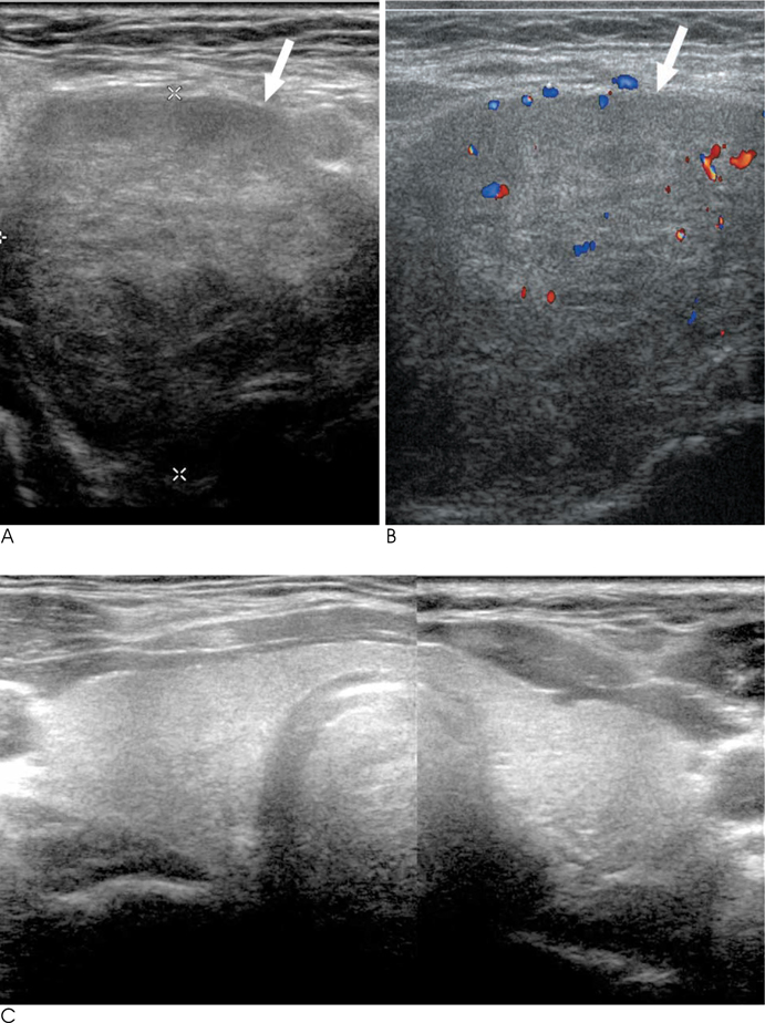
Fig. 3 US findings of the lateral neck mass. A. The gray-scale US shows a circumscribed inhomogeneous mass (arrow) in the left posterior submandibular space. B. The color Doppler scan shows some internal vascularity of the mass (arrow). C. No abnormal lesion is found in the native original thyroid on US.

Fig. 4 Histopathologic findings on the excised mass. A. The photograph of the gross specimen (arrow) shows it is ovoid in shape with a smooth external surface and solid cut surface, and it measures 5.0×3.5 cm. B. Photomicrograph of the specimen shows that the tumor is composed of thyroid tissue (arrow) with small, abortive follicles. The tumor has invaded the vessels (arrowhead) in the outermost portion of the capsule.
Reference
-
1. Mansberger AR Jr, Wei JP. Surgical embryology and anatomy of the thyroid and parathyroid glands. Surg Clin North Am. 1993; 73:727–746.2. Basaria S, Westra WH, Cooper DS. Ectopic lingual thyroid masquerading as thyroid cancer metastases. J Clin Endocrinol Metab. 2001; 86:392–395.3. Subramony C, Baliga M, Lemos LB. Follicular carcinoma arising in ectopic thyroid tissue: case report with fine-needle aspiration findings. Diagn Cytopathol. 1997; 16:39–41.4. Tucci G, Rulli F. Follicular carcinoma in ectopic thyroid gland. A case report. G Chir. 1999; 20:97–99.5. Springer KC. Lingual thyroid: two cases in siblings diagnosed and treated with radioactive iodine. AMA Arch Otolaryngol. 1955; 61:386–393.6. Potdar GG, Desai PB. Carcinoma of the lingual thyroid. Laryngoscope. 1971; 81:427–429.7. Wong KT, Ahuja AT. Ultrasound of thyroid cancer. Cancer Imaging. 2005; 5:157–166.8. Angelos P. Current approaches to the treatment of well-differentiated thyroid cancer. Oncology (Williston Park). 2002; 16:309–331.
- Full Text Links
-
- Actions
-
Cited
- CITED
-
- Close
- Share
-
- Similar articles
-
- Papillary Thyroid Cancer from Lateral Aberrant Thyroid Masquerading as Cervical Metastasis from Larynx Cancer: A Case Report
- A Case of Ectopic Thyroid Papillary Carcinoma with Incidental Papillary Thyroid Microcarcinoma
- Nodular Hyperplasia Arising from the Lateral Aberrant Thyroid Tissue: A Case Report
- A Case of an Ectopic Thyroid Gland at the Lateral Neck Masquerading as a Metastatic Papillary Thyroid Carcinoma
- Adenomatous Hyperplasia Arising from Dual Ectopic Thyroid
