Abdominal Metastasis Arising from Musculoskeletal Sarcoma: CT Findings
- Affiliations
-
- 1Department of Radiology, St.Vincent's Hospital, The Catholic University of Korea, Korea. jeeykim@catholic.ac.kr
- 2Department of Radiology, Seoul St. Mary's Hospital, The Catholic University of Korea, Korea.
- KMID: 2097902
- DOI: http://doi.org/10.3348/jksr.2010.63.3.255
Abstract
- Musculoskeletal sarcoma metastasizes to various sites in the body. The most common metastsis site is the lung, followed less frequently by the skeleton, brain, intra-abdominal organs, lymph node, and pleura. The metastasis of musculoskeletal sarcoma showed various manifestations, which was seen as a nonspecific finding. Metastatic lesions display similar and characteristic features, which include calcification of primary musculoskeletal sarcoma. These features may provide an important diagnostic clue when seen on CT findings. We present a review of the abdominal CT findings in 5 patients with calcified metastasis arising from musculoskeletal sarcoma.
MeSH Terms
Figure
-
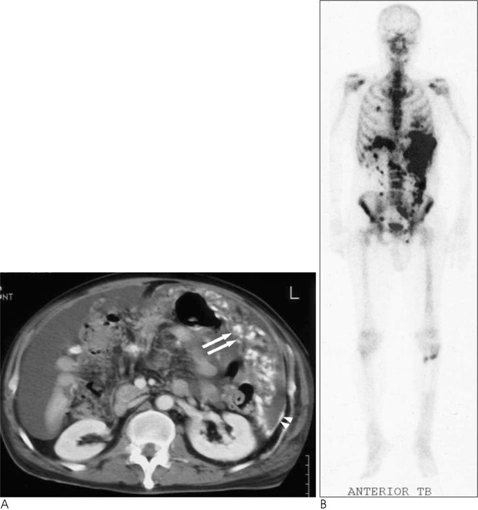
Fig. 1 A 55-year-old man with osteosarcoma. A. Abdominal CT scan demonstrates multiple dense calcified nodules in the omentum (arrows), irregular peritoneal thickening with calcifications (arrowheads), and associated with ascites. B. Scintigraphy with 99mTc-pyrophosphate demonstrates intense uptake in the chest and peritoneum but no evidence of skeletal metastases.

Fig. 2 A 58-year-old female with chondrosarcoma. A, B. Abdominal CT scan demonstrate multiple calcified nodules (arrows) with central arc-like and stippled calcifications in the omentum and mesentery. These nodules show minimal contrast enhancement. C. Abdominal CT scan reveals a soft tissue mass (arrowheads) with chondroid calcifications in left pubic bone.

Fig. 3 A 31-year-old female with osteosarcoma. A, B. Abdominal CT scan show multiple metastatic lymphadenopathy(arrows) with calcifications and strong contrast enhancement in the right external iliac area. C. Plain radiograph demonstrates an ill-defined mass (arrowheads) with cloudlike matrix, cortical break, and extraosseous extension in the head of right fibula.
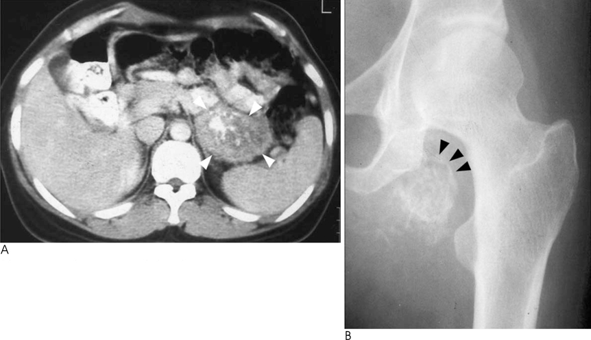
Fig. 4 A 38-year-old woman with juxtacortical chondrosarcoma. A. Abdominal CT reveals a hypodense mass (white arrowheads) with central arc-like and stippled calcifications in the tail of the pancreas. B. Plain radiograph shows a soft tissue mass with stippled calcifications (black arrowheads) in the left thigh with similar chondroid calcifications.
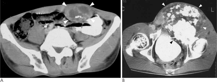
Fig. 5 A 36-year-old man with chondrosarcoma. A. Abdominal CT shows an irregular hypodensemass (arrowheads) with arc-shaped and stippled calcifications in the abdominal wall muscles. B. Abdominal CT scan shows a large mass (arrowheads) with chondroid calcifications in the left pubic bone.
Reference
-
1. Kim SJ, Choi JA, Lee SH, Choi JY, Hong SH, Chung HW, et al. Imaging findings of extrapulmonary metastases of osteosarcoma. Clin Imaging. 2004; 28:291–300.2. Glass RJ, Eftekhari F, Kleinerman ES, Jaffe N, Nachman J. Osteosarcoma metastatic to the pancreas in young patients. Clin Radiol. 1996; 51:293–294.3. Ha HK, Jung JI, Lee MS, Choi BG, Lee MG, Kim YH, et al. CT differentiation of tuberculous peritonitis and peritoneal carcinomatosis. AJR Am J Roentgenol. 1996; 167:743–748.4. Mitchell DG, Hill MC, Hill S, Zaloudek C. Serous carcinoma of the ovary: CT identification of metastatic calcified implants. Radiology. 1986; 158:649–652.5. Pace WM, Ross McDougall I. Tc-99m MDP uptake in soft tissue extraskeletal metastasis from osteogenic sarcoma. Clin Nucl Med. 2000; 25:333–334.6. Jeffree GM, Price CH, Sissons HA. The metastatic patterns of osteosarcoma. Br J Cancer. 1975; 32:87–107.7. van Zanten TE, Golding RP, Taets ven Amerongen AH. Osteosarcoma with calcific mediastinal lymphadenopathy. Pediatr Radiol. 1987; 17:258–259.8. Buetow PC, Parrino TV, Buck JL, Pantongrag-Brown L, Ros PR, Dachman AH, et al. Islet cell tumors of the pancreas: pathologic-imaging correlation among size, necrosis and cysts, calcification, malignant behavior, and functional status. AJR Am J Roentgenol. 1995; 165:1175–1179.9. Buetow PC, Buck JL, Pantongrag-Brown L, Beck KG, Ros PR, Adair CF. Solid and papillary epithelial neoplasm of the pancreas: imaging-pathologic correlation on 56 cases. Radiology. 1996; 199:707–711.10. Klein KA, Stephens DH, Welch TJ. CT characteristics of metastatic disease of the pancreas. Radiographics. 1998; 18:369–378.
- Full Text Links
-
- Actions
-
Cited
- CITED
-
- Close
- Share
-
- Similar articles
-
- CT and Positron Emission Tomography/CT Findings of Mediastinal Extraskeletal Ewing's Sarcoma with Extensive Distant Metastasis: A Case Report
- A Case of Epithelioid Sarcoma in a Child
- CT Findings of Primary Undifferentiated Pleomorphic Sarcoma in the Small Bowel: A Case Report
- Alveolar soft-part sarcoma
- A Case of Clear Cell Sarcoma Developing Early Systemic Metastasis
