J Korean Soc Radiol.
2010 Sep;63(3):221-224. 10.3348/jksr.2010.63.3.221.
Thymic Carcinoma Presenting Two Independent Nodules: Case Report
- Affiliations
-
- 1Department of Radiology, Korea University Hospital, Korea University College of Medicine, Korea. kiylee@korea.ac.kr
- 2Department of Internal Medicine, Korea University Hospital, Korea University College of Medicine, Korea.
- KMID: 2097896
- DOI: http://doi.org/10.3348/jksr.2010.63.3.221
Abstract
- Thymic carcinoma is a rare malignant neoplasm of the thymus arising in the thymic epithelium and has a higher frequency of local invasion and metastasis than other subtypes of thymic epithelial tumors. Thymic carcinoma is usually demonstrated as a large, irregular mass located in the anterior mediastinum and commonly contain a necrotic or cystic component. We report atypical CT findings and multicentricity in a case of thymic carcinoma presenting two small nodules in the anterior mediastinum.
Figure
-

Fig. 1 Chest radiograph PA projection shows no definite abnormal findings.
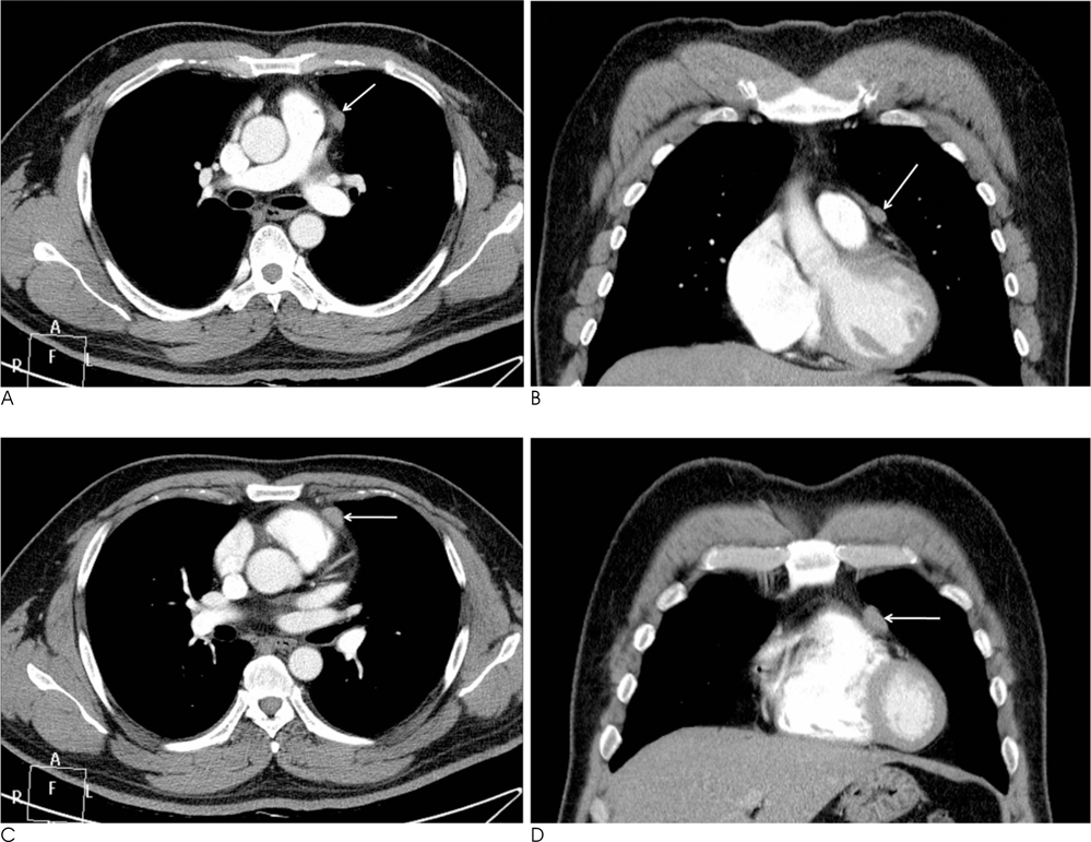
Fig. 2 Axial CT images show two well defined nodules (arrows) abutting pericardial fat in left anterior mediastinum at the level of main bronchus (Fig. 2A, B) and upper right ventricle (Fig. 2C, D).
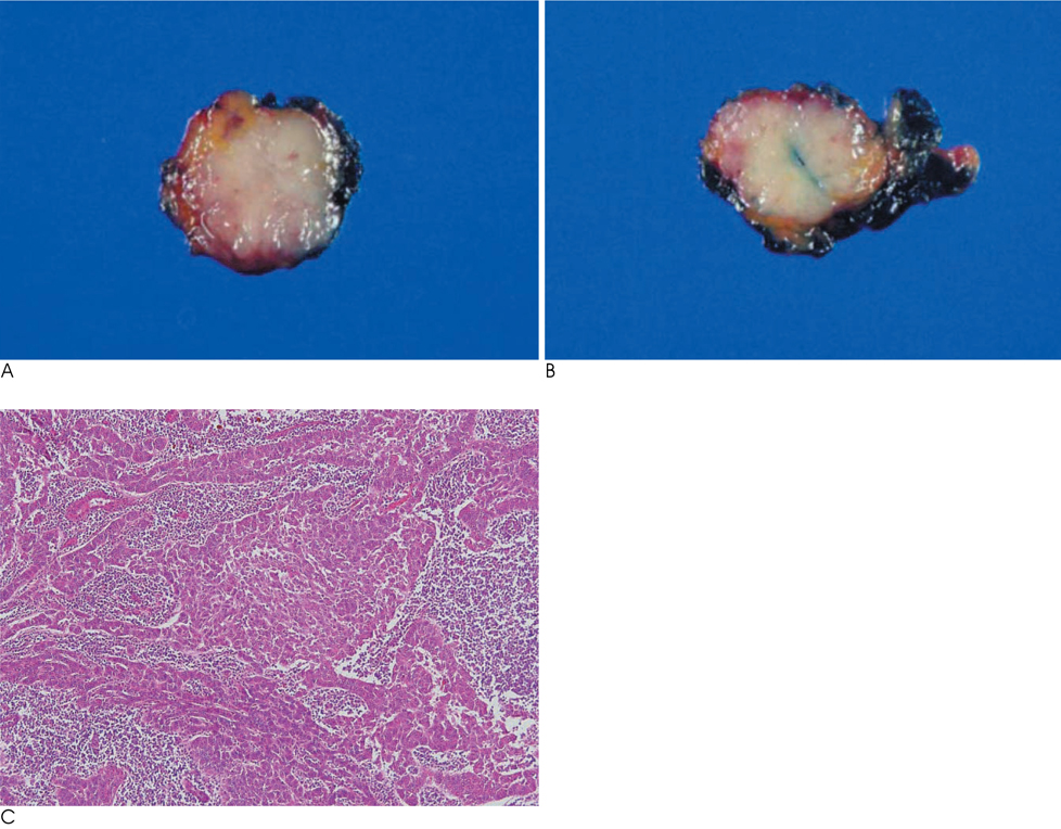
Fig. 3 A, B. Cut surface of the two nodules show well demarcated, whitish gray solid appearance. C. Photomicrograph (H & E, ×100) shows epithelial cells with severe nuclear atypia, mitotic figures and parenchymal invasion of pleomorphic tumor cell nests.
Reference
-
1. Eng TY, Fuller CD, Jagirdar J, Bains Y, Thomas CR Jr. Thymic carcinoma: state of the art review. Int J Radiat Oncol Biol Phys. 2004; 59:654–664.2. Okumura M, Ohta M, Tateyama H, Nakagawa K, Matsumura A, Maeda H, et al. The World Health Organization histologic classification system reflects the oncologic behavior of thymoma: a clinical study of 273 patients. Cancer. 2002; 94:624–632.3. Jeong YJ, Lee KS, Kim J, Shim YM, Han J, Kwon OJ. Does CT of thymic epithelial tumors enable us to differentiate histologic subtypes and predict prognosis? AJR Am J Roentgenol. 2004; 183:283–289.4. Sadohara J, Fujimoto K, Muller NL, Kato S, Takamori S, Ohkuma K, et al. Thymic epithelial tumors: comparison of CT and MR imaging findings of low-risk thymomas, high-risk thymomas, and thymic carcinomas. Eur J Radiol. 2006; 60:70–79.5. Jung KJ, Lee KS, Han J, Kim J, Kim TS, Kim EA. Malignant thymic epithelial tumors: CT-pathologic correlation. AJR Am J Roentgenol. 2001; 176:433–439.6. Nomori H, Kobayashi K, Ishihara T, Suito T, Torikata C. A case of multiple thymoma: the possibility of intra-thymic metastasis. Jpn J Clin Oncol. 1990; 20:209–211.7. Nonami Y, Moriki T. Synchronous independent bifocal orthotopic thymomas. A case report. J Cardiovasc Surg. 2004; 45:585–587.8. Yoneda S, Matsuzoe D, Kawakami T, Tashiro Y, Shirahama H, Ohkubo K, et al. Synchronous multicentric thymona: report of a case. Surg Today. 2004; 34:597–599.9. Kawaguchi K, Usami N, Uchiyama M, Ito S, Yasuda A, Yokoi K. Triple thymoma with different histologic types. J Thorac Cardiovasc Surg. 2007; 133:826–827.10. Lucchi M, Mussi A, Basolo F, Ambrogi MC, Fontanini G, Angeletti CA. The multimodality treatment of thymic carcinoma. Eur J Cardiothorac Surg. 2001; 19:566–569.
- Full Text Links
-
- Actions
-
Cited
- CITED
-
- Close
- Share
-
- Similar articles
-
- Postoperative Radiotherapy in Thymic Carcinoma : A case report
- A Case of Intracardiac Thymic Carcinoma Presenting as Congestive Hepatopathy
- A Case of Thymic Carcinoma with Direct Invasion into the Skin
- A Case of Cutaneous Matestasis Originating from Thymic Carcinoma
- A Case of Well-Differentiated Thymic Carcinoma with Extensive Cystic Degeneration
