A Change of Pulmonary Function in the Patients with Severe Non-Idiopathic Scoliosis Following Reconstructive Spine Surgery
- Affiliations
-
- 1Department of Orthopedic Surgery, Yonsei University College of Medicine, Korea. mes1007@yumc.yonsei.ac.kr
- 2Department of Orthopaedic Surgery, National Health Insurance Corporation Ilsan Hospital, Korea.
- 3Department of Orthopaedic Surgery, Cha University College of Medicine, Korea.
- 4Department of Orthopaedic Surgery, Ahn Sei Hospital, Korea.
- KMID: 2097826
- DOI: http://doi.org/10.4184/jkss.2006.13.2.106
Abstract
-
STUDY DESIGN: This is a retrospective study
OBJECTIVES
We wanted to investigate the changes in pulmonary function after spine surgery for the patients suffering with severe non-idiopathic scoliosis. SUMMARY OF LITERATURE REVIEW: The potential for pulmonary function change after scoliosis surgery may be much greater for the patients with neuromuscular, congenital or neuro-fibromatosis because of the severe thoracic deformity. Yet few studies have been performed on this subject.
MATERIALS AND METHODS
12 non-idiopathic scoliosis patients (average age: 11.1 years) were followed up for more than one year. Among these patients, 7 had muscular dystrophy, 3 had spinal muscular atrophy, 1 had Guillain-BarreSyndrome and 1 had neuro-fibromatosis. Surgery was done through the anterior and posterior approaches in 7 cases, and the posterior approach was used in 5 cases. In the former group, open thoracotomy was performed in 6 cases. NIPPV (non-invasive positive pressure ventilation) was used for 4 patients before surgery.
RESULTS
The average Cobb's angle improved from preoperative 93.9 to postoperative 42.4, showing 55% correction. The average FVC was 1270ml before surgery and 1365 ml postoperatively, and the average FEV1 was 1163 ml preoperatively and 1300 ml postoperatively, showing a slightly increased FEV1. When these data were analyzed in detail, the FVC was decreased from preoperative 72.3% to 63.8% postoperatively in the 6 patients who underwent open thoracotomy. On the other hand, it was increased from preoperative 43% to 47.5% postoperatively in the 6 patients who did not undergo thoracotomy.
CONCLUSION
Although the pulmonary function deteriorated after reconstructive spine surgery in some patients, worsening was seen mainly in those patients who underwent thoracotomy. The pulmonary function was actually improved in the patients who underwent surgery without thoracotomy to correct their severe scoliosis
Keyword
MeSH Terms
Figure
-
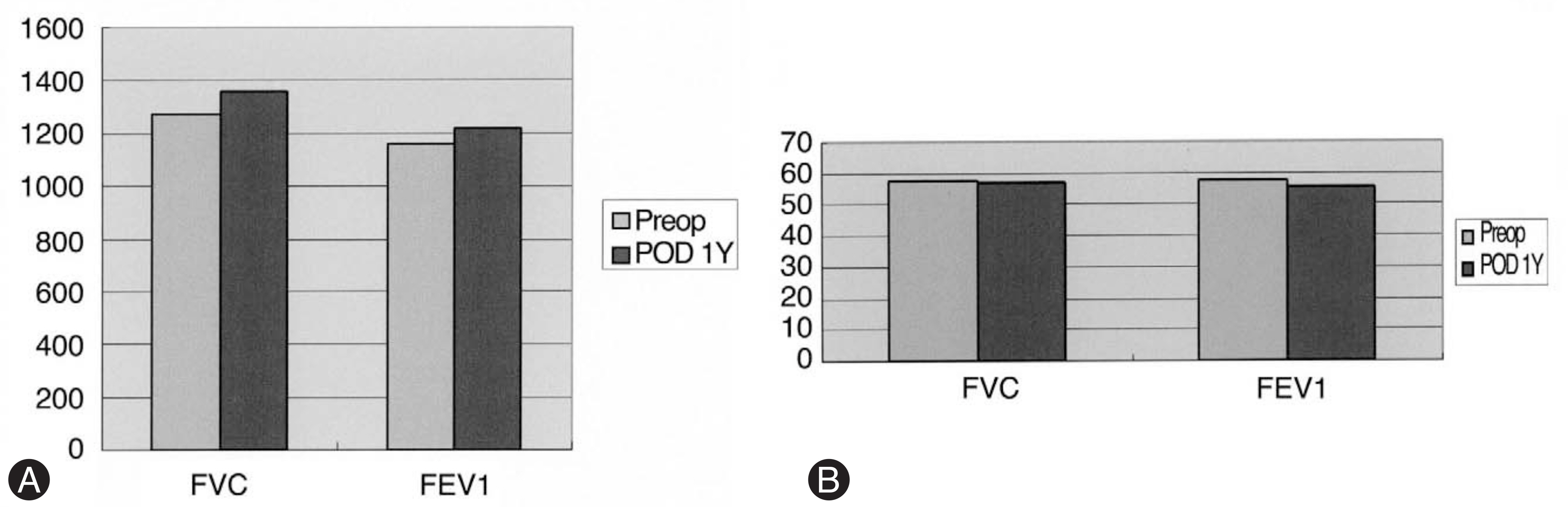
Fig. 1. The change of the FVC and FEV1 in the volume (A) and the percentage of predicted normal values(B). (A) The average volume of FVC was 1270 ml before surgery and 1365 ml by postoperative 1 year, and the average FEV1 was 1163 ml and 1300 ml, showing slightly increase in FEV1. (B) The average percentage of FVC decreased from 57.3% before surgery to 56.6% by postoperative 1yr and the average FEV1 was also decreased from 57.3% to 55.2%.
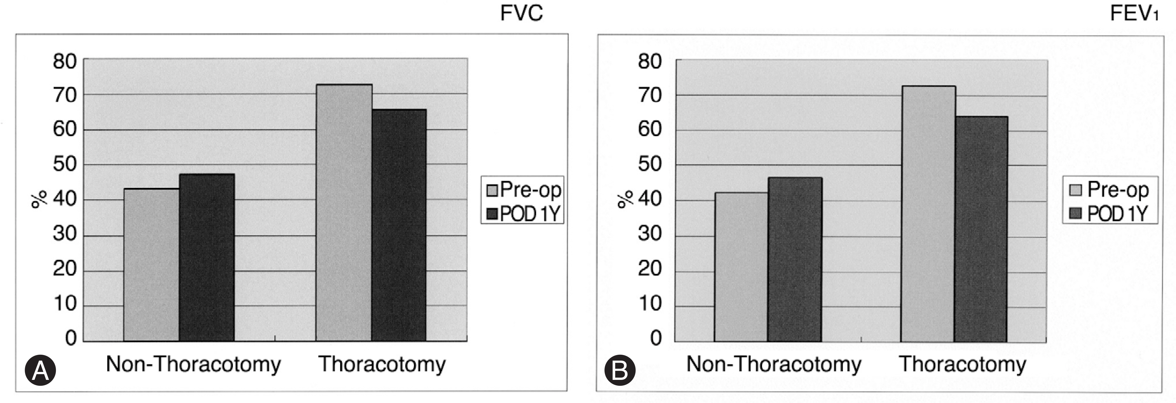
Fig. 2. When these data were analyzed in detail, FVC (A) was decreased from preoperative 72.3% to 63.8% postoperatively in 6 patients who underwent open thoracotomy. On the other hand, it was increased from preoperative 43% to 47.5% post-operative 1year in 6 patients. FEV1 (B) was decreased from preoperative 72.5% to 63.8% postoperatively in 6 patients who underwent open thoracotomy. On the other hand, it was increased from preoperative 42.2% to 46.5% postoperative 1year in 6 patients.

Fig. 3. Case 11. (A, B) Sitting posteroanterior (PA) and lateral radiography of 12 year 5 month patient with spinal muscular atrophy. (C, D) Postoperative sitting PA and lateral radiography. The clinical picture shows (E) evident pelvic obliquity and (F) the correction of pelvic obliquity after operation.
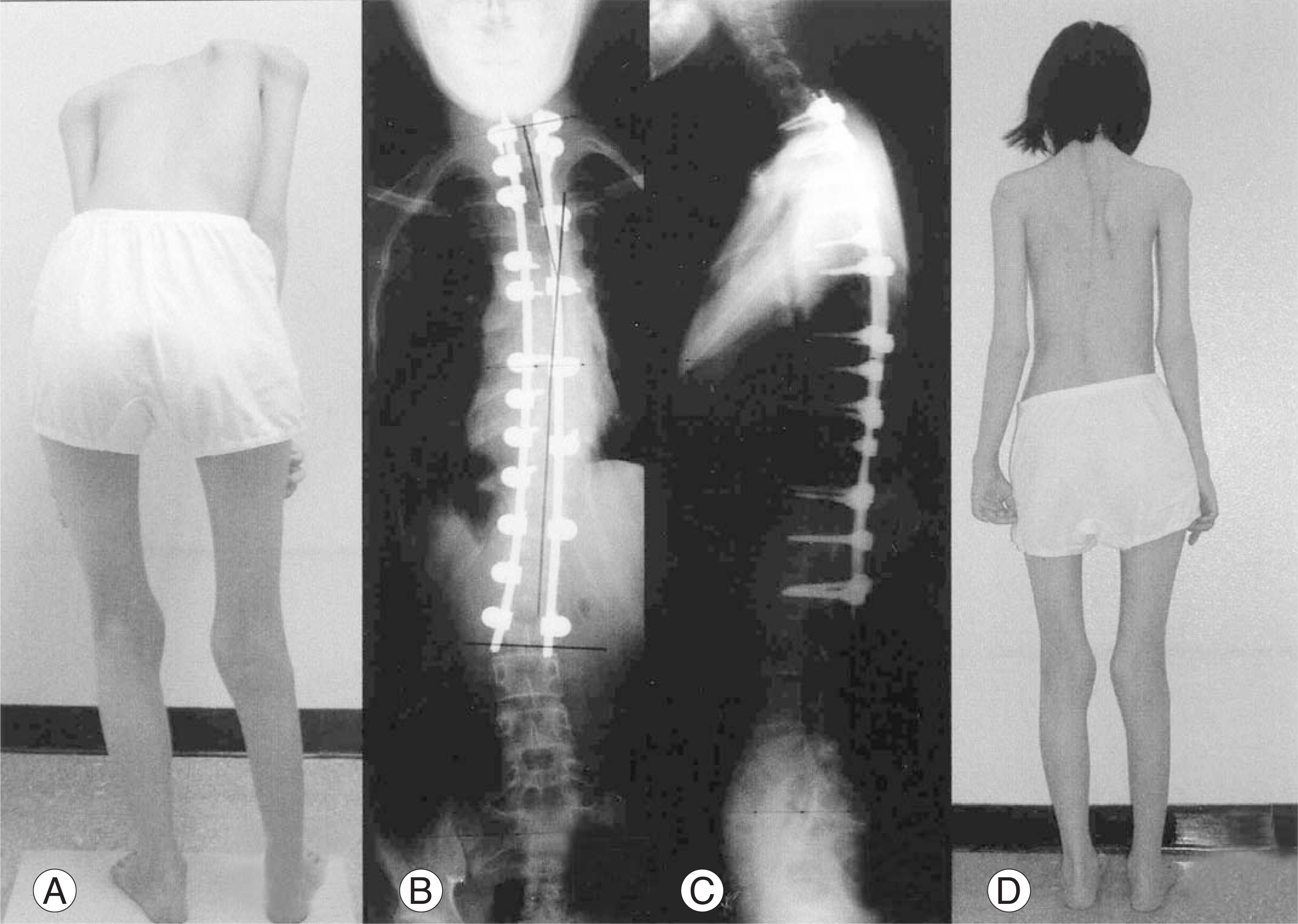
Fig. 4. Preoperative picture (A) of a 13-yr-old Guillain Barre syndrome patient. Before operation, she can stand for a moment. Post-operative PA (B) and lateral (C) radiography with pedicle technique the scoliosis was reduced to less than 20 degrees. (D) She can walk without support after opeatrion
Reference
-
01). Bridwell KH. Surgical treatment of idiopathic adolescent scoliosis. Spine. 1999. 24:2607–2616.
Article02). Wazeka AN., DiMaio MF., Boachie-Adjei O. Outcome of pediatric patients with sevre restrictive lung disease following reconstructive spine surgery. Spine. 2004. 29:528–535.03). Kinnear WJM., Johnston IDA. Does Harrington instrumentation improve ulmonary function in adolescents with idiopathic scoliosis? Spine. 1993. 18:1556–1559.04). Lamarre A., Hall JE., Weng TR., Aspin N., Levison H. Pulmonary function in Scoliosis one year after surgical correction. Proceedings of the Scoliosis Research Society. J Bone Joint Surg [Am]. 1971. 53:195.05). Schneerson JM., Edgar MA. Cardiac and respiratory function before and after spinal arthrodesis in adolescent idiopathic scoliosis. Thorax. 1979. 34:658–661.06). Suk SI., Lee CS., Yoon GS. Changes of Pulmonary Function after Surgical Correction in Scoliosis. J Kor Orthop Assoc. 1984. 19:1067–1072.
Article07). Lenke LG., Bridwell KH., Baldus C., Blanke K. Analysis of pulmonary patients treated with Cotrel-Dubousset instrumentation. J Spinal Disord. 1992. 5:16–25.08). Shufflebarger HL., Gepstein R., Clark C. Recovery of pulmonary function after Cotrel-Dubousset instrumentation (CDI). Orthop Trans. 1987. 11:498–499.09). Wood KB., Schendel MJ., Dekutoski MB., Boachie-Adjei O., Heithoff KH. Thoracic volume changes in scoliosis surgery. Spine. 1996. 21:718–723.
Article10). Kumano K., Tsuyama N. Pulmonary function before and after surgical correction of Scoliosis. J Bone Joint Surg [Am]. 1982. 64:242–248.
Article11). American Thoracic Society. Lung function testing: selection of reference values and interpretative strategies. Am Rev Respir Dis. 1991. 144:1202–1218.12). Jones P., Quirk F., and Baveystock C. The St George's Respiratory Questionnaire. Respiratory Medicine. 1991. 85sup B:25–31.
Article13). Kennedy JD., Staples AJ., Brook PD, et al. Effect of spinal surgery on lung function in Duchenne muscular dystrophy. Thorax. 1995. 50:1173–1178.
Article14). Kurz LT., Mubarak SJ., Schultz P., Park SM., Leach J. Correlation of scoliosis and pulmonary function in Duchenne muscular dystrophy. J Pediatr Orthop. 1983. 3:347–353.
Article15). Galasko CS., Delaney C., Morris P. Spinal stabilization in Duchenne muscular dystrophy. J Bone Joint Surg [Br]. 1992. 74:210–214.16). Thacker M., Hui JHP., Wong HK., Chatterjee A., Lee EH. Spinal fusion and instrumentation for paediatric neuromuscular scoliosis: retrospective review. J Orthop Surg (Hong Kong). 2002. 10:144–151.
Article17). Chng SY., Wong YQ., Hui JH., Wong HK., Ong HT., Goh DY. Pulmonary function and scoliosis in children with spinal muscular atrophy types II and III. J Paediatr Child Health. 2003. 39:673–676.
Article18). Vedantam R., Lenke LG., Bridwell KH., Haas J., Linville DA. A prospective evaluation of pulmonary function in patients with adolescent idiopathic scoliosis relative to the surgical approach used for spinal arthrodesis. Spine. 2000. 25:82–90.
Article19). Wong CA., Cole AA., Watson L., Webb JK., Johnston ID., Kinnear WJ. Pulmonary function before and after anterior spinal surgery in adult idiopathic scoliosis. Tho- rax. 1996. 51:1556–1559.
Article20). Graham EJ. Lenke LG., Lowe TG, et al. Prospective pulmonary function evaluation following open thoracoto my for anterior spinal fusion in adolescent idiopathic scoliosis. Spine. 2000. 25:2319–2325.
