J Korean Soc Spine Surg.
2001 Jun;8(2):176-180. 10.4184/jkss.2001.8.2.176.
Dysphagia Caused by an Anterior Cervical Osteophyte: A Case Report
- Affiliations
-
- 1Department of Orthopaedic Surgery, College of Medicine, Pusan National University, Pusan, Korea. pww@scoliosis.co.kr
- KMID: 2097759
- DOI: http://doi.org/10.4184/jkss.2001.8.2.176
Abstract
-
INTRODUCTION: The dysphagia due to the osteophyte of the anterior cervical spine was reported to occur in the old age and lower cervical spine. The authors experienced a case of dysphagia associated with osteophyte of the 3-4th anterior cervical spine without trauma history in a young man.
MATERIALS AND METHODS
A 37-year-old man presented with the dysphagia during 2 months. The lateral radiograph and com-puted tomograph of cervical spine showed 1.5 cm sized anterior osteophyte in the 3-4th cervical vertebrae that compressed the arytenoid cartilage anteriorly. In the finding of video laryngoscopy and endoscopy, the posterior hypopharyngeal wall was pro-truded and reflex gastritis was also seen. Through the anterior approach, the excision of osteophyte, discectomy and bone graft was done. RESULT: Dyaphagia was relieved immediately after the removal of osteophyte. The follow up video laryngoscopy showed that the posterior hypopharyngeal wall was normalized. The radiograph showed bone union and no recurrence at 3 year follow up.
CONCLUSION
The surgical removal of anterior cervical osteophyte causing dysphasia showed symptom relief and excellent result at the long-term follow up.
Keyword
MeSH Terms
Figure
-

Fig. 1. Telescopic video laryngoscopy demonstrating the bulging out of posterior hypopharyngeal wall.

Fig. 2. Lateral radiography of cervical spine. It shows protruding large osteophyte at C3-4 vertebrae.
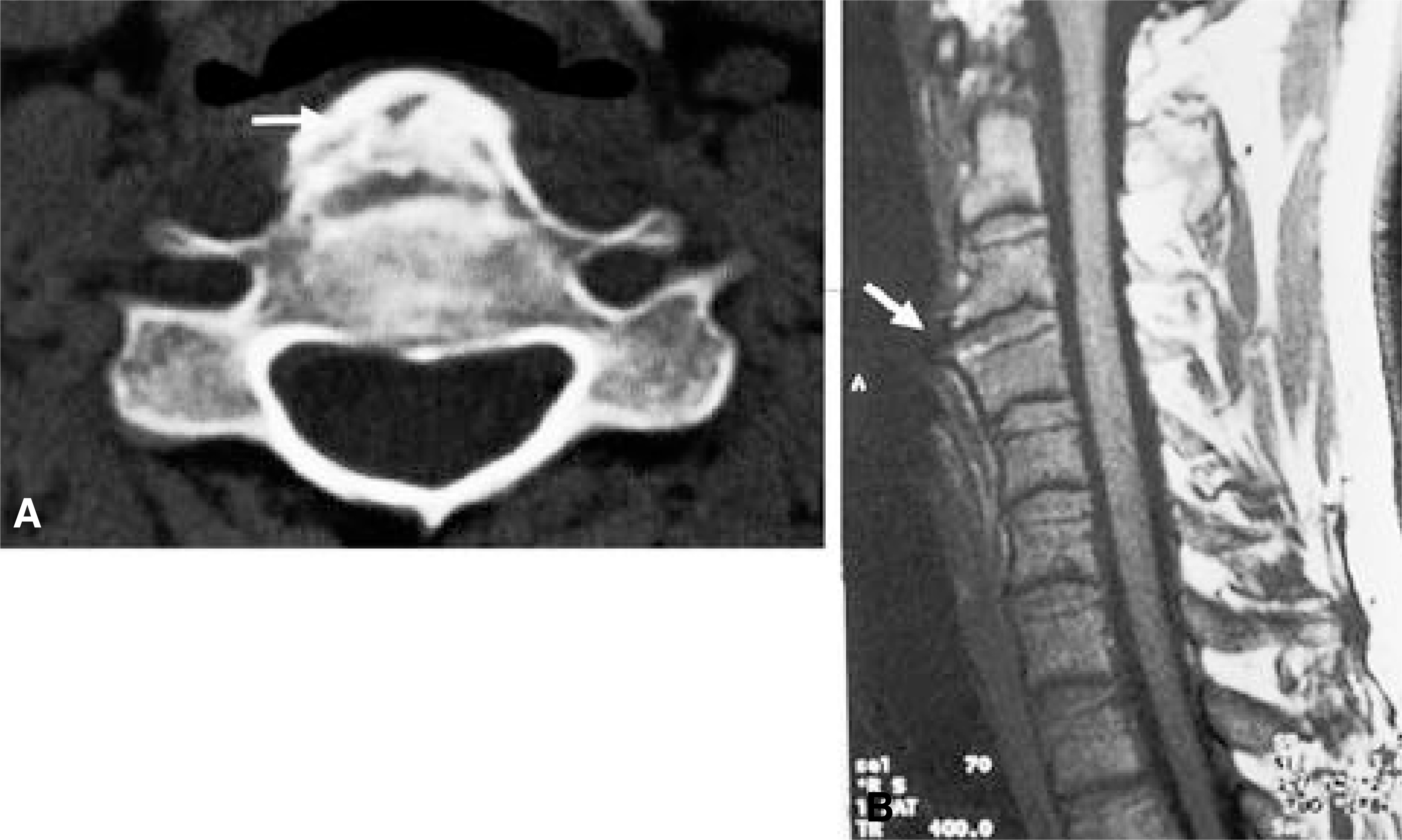
Fig. 3-A. CT scan at the level of hypopharynx demonstrates that abnormal osteophyte displaces and compress the posterior hypopharyngeal wall. Fig. 3-B. MRI finding shows protruding osteophyte but normal intervertebral disc space.

Fig. 4-A. Lateral radiography of cervical spine after excision of osteophyte, discectomy and bone graft. Fig. 4-B. Telescopic video laryngoscopy shows normal posterior hypopharyngeal wall.

Fig. 5. Postoperative 3 year lateral radiograph shows solid bone union between C3-4 vertebral body.
Reference
-
1). Baek CH, Kim SM, Chu KC. A Case of Diffuse Idiopathic Skeletal Hyperostosis with Dysphagia, Korean J Otolaryngol. 41:1498–1496. 1998.2). Burkus JK. Esophageal Obstruction Secondary to Diffuse Idiopathic Skeletal Hyperostosis, Orthopedics. 11:717–720. 1988.3). Davies RP, Sage MR. Cervical osteophyte Induced dysphagia, Australasian Radiology. 33:223–225. 1989.4). Demuynck K, Calenburgh FV, Goffin J, Verschakelen J, Demedts M, Woestijne KV. Upper airway obstruction caused by a cervical osteophyte. Chest. 108:283–284. 1995.
Article5). Forestier J, Ragier R. Ankylosing hyperostosis of the spine. Clin Orthop. 74:65–83. 1971.
Article6). Gamach FW, Voorhies RM. Hypertrophic cervical osteophytes causing dysphagia. J Neurosurg. 53:338–344. 1980.7). Hirano H, Suzuki H, Sakakibara T, Higuchi Y, Inoue K, Suzuki Y. Dysphagia due to hypertrophic cervical osteophytes. Clin Orthop. 162:168–172. 1982.
Article8). Kim KC, Youn SH, Park HS, et al. Dysphagia due to Cervical Osteophytes: Case Report. J Korean Neurosurg. 27:109–113. 1998.9). Kissel P, Youmans JR. Posttraumatic anterior cervical osteophyte and dysphagia: surgical report and literature review. Journal of Spinal Disorders. 5:104. 1992.10). Kodama M, Sawada H, Udaka F, Kameyama M, Koyama T. Dysphagia caused by an anterior cervical osteophyte: case report. Neuroradiology. 37:58. 1995.
Article11). Maran A, Jacobson I. Cervical osteophytes presenting with pharyngeal symptoms. Laryngoscope. 81:412–417. 1971.
Article12). Marra A, Dario A, Scamoni C, Pozzi M, Soldati M, Dorizzi A. Dysphagia due to anterior cervical osteophyte. J Neurosurg Sci. 35:229–231. 1991.13). McGarrah PD, Teller D. Posttraumatic cervical osteophytosis causing progressive dysphagia. Southern Medical Journal. 90:858–860. 1997.
Article
- Full Text Links
-
- Actions
-
Cited
- CITED
-
- Close
- Share
-
- Similar articles
-
- Temporarily Aggravated Dysphagia Following Osteophytectomy and Fixation in a Patient with Cervical Osteophyte
- A rare cause of dysphagia: compression of the esophagus by an anterior cervical osteophyte due to ankylosing spondylitis
- Giant Anterior Cervical Osteophyte Leading to Dysphagia
- Anterior Herniation of Partially Calcified and Degenerated Cervical Disc Causing Dysphagia
- Improvement of Dysphagia after Anterior Cervical Screw Removal: Case Report
