J Lipid Atheroscler.
2013 Jun;2(1):9-17. 10.12997/jla.2013.2.1.9.
Hypertension and Increased Left Ventricular End-diastolic Pressure Influence Arterial Stiffness
- Affiliations
-
- 1Division of Cardiology, Department of Internal Medicine, Gachon University, Gil Hospital, Incheon, Korea. msshin@gilhospital.com
- 2Department of Internal Medicine, Seoul Medical Center, Seoul, Korea.
- KMID: 2095090
- DOI: http://doi.org/10.12997/jla.2013.2.1.9
Abstract
OBJECTIVE
There have been few studies regarding the relationship between arterial stiffness and left ventricular end-diastolic pressure (LVEDP). In the current study, we evaluated the relationship between the LVEDP and arterial stiffness in patients with hypertension (HTN).
METHODS
Group I (n=34) included patients with a normal E/E' (< or =8) without HTN, group II (n=31) included patients with an elevated E/E' (>8) without HTN, group III (n=20) included patients with a normal E/E' (< or =8) with HTN, and group IV (n=49) included patients with an elevated E/E' (>8) with HTN. Aortic distensibility (AD) and the right brachial-ankle pulse wave velocity (baPWV) were measured.
RESULTS
The mean age was 46.0+/-11.3 years. The mean value of AD was significantly lower in the group III compared to the group I. The group IV showed significantly lower AD compared to the group II. The group III demonstrated higher baPWV compared to the group I (1422+/-182 cm/sec vs. 1186+/-178 cm/sec, p<0.01), and the group IV showed higher baPWV compared to the group II (1456+/-228 vs. 1259+/-238 cm/sec, p<0.01). However, AD and baPWV were not significantly different between the group I and II, and between the group III and IV. The E/E' ratio showed a weak negative correlation with AD and a weak positive correlation with baPWV.
CONCLUSION
Patients with hypertension showed a lower AD and a higher baPWV compared to those with normal blood pressure independent of the LVEDP. But the correlation between E/E' ratio and arterial stiffness suggests that a high LVEDP might not significantly influence arterial stiffness.
Figure
-
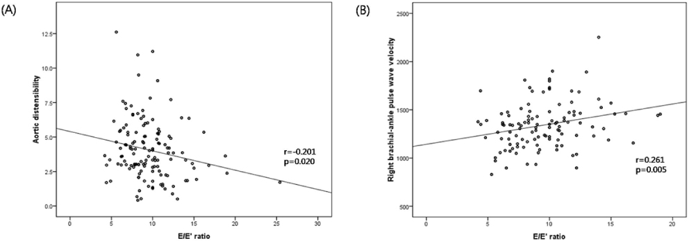
Fig. 1 Correlation of E/E' ratio and arterial stiffness (aortic distensibility (A), brachial-ankle pulse wave velocity (B))

Fig. 2 Correlation of brachial-ankle pulse wave velocity and aortic distensibility in lower left ventricular end-diastolic pressure (LVEDP) group (group I and III) (A), and higher LVEDP group (group II and IV) (B).
Reference
-
1. Avolio AP, Chen SG, Wang RP, Zhang CL, Li MF, O'Rourke MF. Effects of aging on changing arterial compliance and left ventricular load in a northern Chinese urban community. Circulation. 1983; 68:50–58.
Article2. Lehmann ED, Gosling RG, Sönksen PH. Arterial wall compliance in diabetes. Diabet Med. 1992; 9:114–119.
Article3. Wada T, Kodaira K, Fujishiro K, Maie K, Tsukiyama E, Fukumoto T, Uchida T, Yamazaki S. Correlation of ultrasound-measured common carotid artery stiffness with pathological findings. Arterioscler Thromb. 1994; 14:479–482.
Article4. London GM, Marchais SJ, Safar ME, Genest AF, Guerin AP, Metivier F, Chedid K, London AM. Aortic and large artery compliance in end-stage renal failure. Kidney Int. 1990; 37:137–142.
Article5. Wang CP, Hung WC, Yu TH, Hsu HL, Chen YH, Chiu CA, Lu LF, Chung FM, Cheng YA, Lee YJ. Brachial-ankle pulse wave velocity as an early indicator of left ventricular diastolic function among hypertensive subjects. Clin Exp Hypertens. 2009; 31:31–43.
Article6. Devereux RB, Savage DD, Sachs I, Laragh JH. Relation of hemodynamic load to left ventricular hypertrophy and performance in hypertension. Am J Cardiol. 1983; 51:171–176.
Article7. Bouthier JD, De Luca N, Safar ME, Simon AC. Cardiac hypertrophy and arterial distensibility in essential hypertension. Am Heart J. 1985; 109:1345–1352.
Article8. Lartaud-Idjouadiene I, Lompré AM, Kieffer P, Colas T, Atkinson J. Cardiac consequences of prolonged exposure to an isolated increase in aortic stiffness. Hypertension. 1999; 34:63–69.
Article9. Eren M, Gorgulu S, Uslu N, Celik S, Dagdeviren B, Tezel T. Relation between aortic stiffness and left ventricular diastolic function in patients with hypertension, diabetes, or both. Heart. 2004; 90:37–43.
Article10. Blacher J, Asmar R, Djane S, London GM, Safar ME. Aortic pulse wave velocity as a marker of cardiovascular risk in hypertensive patients. Hypertension. 1999; 33:1111–1117.
Article11. Yamashina A, Tomiyama H, Arai T, Hirose K, Koji Y, Hirayama Y, Yamamoto Y, Hori S. Brachial-ankle pulse wave velocity as a marker of atherosclerotic vascular damage and cardiovascular risk. Hypertens Res. 2003; 26:615–622.
Article12. Sahn DJ, DeMaria A, Kisslo J, Weyman A. Recommendations regarding quantitation in M-mode echocardiography: results of a survey of echocardiographic measurements. Circulation. 1978; 58:1072–1083.
Article13. Devereux RB, Reichek N. Echocardiographic determination of left ventricular mass in man. Anatomic validation of the method. Circulation. 1977; 55:613–618.
Article14. Karamitsos TD, Karvounis HI, Didangelos TP, Papadopoulos CE, Kachrimanidou MK, Selvanayagam JB, Parharidis GE. Aortic elastic properties are related to left ventricular diastolic function in patients with type 1 diabetes mellitus. Cardiology. 2008; 109:99–104.
Article15. Tsioufis C, Chatzis D, Dimitriadis K, Stougianos P, Kakavas A, Vlasseros I, Tousoulis D, Stefanadis C, Kallikazaros I. Left ventricular diastolic dysfunction is accompanied by increased aortic stiffness in the early stages of essential hypertension: a TDI approach. J Hypertens. 2005; 23:1745–1750.
Article16. Abhayaratna WP, Srikusalanukul W, Budge MM. Aortic stiffness for the detection of preclinical left ventricular diastolic dysfunction: pulse wave velocity versus pulse pressure. J Hypertens. 2008; 26:758–764.
Article17. Malayeri AA, Natori S, Bahrami H, Bertoni AG, Kronmal R, Lima JA, Bluemke DA. Relation of aortic wall thickness and distensibility to cardiovascular risk factors (from the Multi-Ethnic Study of Atherosclerosis [MESA]). Am J Cardiol. 2008; 102:491–496.
Article18. Triposkiadis F, Kallikazaros I, Trikas A, Stefanadis C, Stratos C, Tsekoura D, Toutouzas P. A comparative study of the effect of coronary artery disease on ascending and abdominal aorta distensibility and pulse wave velocity. Acta Cardiol. 1993; 48:221–233.19. Stefanadis C, Stratos C, Boudoulas H, Kourouklis C, Toutouzas P. Distensibility of the ascending aorta: comparison of invasive and non-invasive techniques in healthy men and in men with coronary artery disease. Eur Heart J. 1990; 11:990–996.
Article20. Montalcini T, Gorgone G, Pujia A. Association between pulse pressure and subclinical carotid atherosclerosis in normotensive and hypertensive post-menopausal women. Clin Exp Hypertens. 2009; 31:64–70.
Article21. Koji Y, Tomiyama H, Ichihashi H, Nagae T, Tanaka N, Takazawa K, Ishimaru S, Yamashina A. Comparison of ankle-brachial pressure index and pulse wave velocity as markers of the presence of coronary artery disease in subjects with a high risk of atherosclerotic cardiovascular disease. Am J Cardiol. 2004; 94:868–872.
Article22. Nagueh SF, Middleton KJ, Kopelen HA, Zoghbi WA, Quiñones MA. Doppler tissue imaging: a noninvasive technique for evaluation of left ventricular relaxation and estimation of filling pressures. J Am Coll Cardiol. 1997; 30:1527–1533.
Article23. Nagueh SF, Mikati I, Kopelen HA, Middleton KJ, Quiñones MA, Zoghbi WA. Doppler estimation of left ventricular filling pressure in sinus tachycardia. A new application of tissue doppler imaging. Circulation. 1998; 98:1644–1650.
Article24. Firstenberg MS, Levine BD, Garcia MJ, Greenberg NL, Cardon L, Morehead AJ, Zuckerman J, Thomas JD. Relationship of echocardiographic indices to pulmonary capillary wedge pressures in healthy volunteers. J Am Coll Cardiol. 2000; 36:1664–1669.
Article25. On YK, Kim MK, Jeung HS, Hyun MS, Kim SK, Kwon YJ. Echocardiographic indices associated with left ventricular end-diastolic pressure. Korean Circ J. 2002; 32:872–877.
Article26. Liang HY, Cauduro SA, Pellikka PA, Bailey KR, Grossardt BR, Yang EH, Rihal C, Seward JB, Miller FA, Abraham TP. Comparison of usefulness of echocardiographic Doppler variables to left ventricular end-diastolic pressure in predicting future heart failure events. Am J Cardiol. 2006; 97:866–871.
Article
- Full Text Links
-
- Actions
-
Cited
- CITED
-
- Close
- Share
-
- Similar articles
-
- The Influence of the Left Ventricular Geometry on the Left Atrial Size and Left Ventricular Filling Pressure in Hypertensive Patients, as Assessed by Echocardiography
- Effect of Atenolol on Left Ventricular Function in Essential Hypertension
- Short-term Treatment with Angiotensin II Antagonist in Essential Hypertension:Effects of Losartan on Left Ventricular Diastolic Function, Left Ventricular Mass, and Aortic Stiffness
- Left ventricular hypertrophy and diastolic function in children and adolescents with essential hypertension
- Correlations between the Left Ventricular Diastolic Function and Aortic Stiffness in Healthy Aged Subjects
