Cystic Papillary Renal Cell Carcinoma Arising from an Involutional Multicystic Dysplastic Kidney
- Affiliations
-
- 1Department of Diagnostic Radiology, Jeju National University School of Medicine, Jeju National University Hospital, Jeju, Korea. 67kbs@medimail.co.kr
- 2Department of Urology, Jeju National University School of Medicine, Jeju National University Hospital, Jeju, Korea.
- 3Department of Pathology, Jeju National University School of Medicine, Jeju National University Hospital, Jeju, Korea.
- KMID: 2079571
- DOI: http://doi.org/10.3348/jksr.2015.73.5.343
Abstract
- Multicystic dysplastic kidney is a common cystic renal disease that often occurs in infancy. Recent studies demonstrate the possibility for spontaneous involution of a dysplastic kidney. In such cases, the prognosis is generally excellent and there is a very low incidence of complications. Complications associated with multicystic dysplastic kidney include pain, infection, hypertension, and neoplasia. Renal cell carcinomas are extremely rare in multicystic dysplastic kidneys. To our knowledge, no case report has described a radiologic finding of renal cell carcinoma arising from an involutional multicystic dysplastic kidney. We report a case of histopathologically validated cystic papillary renal cell carcinoma arising from an involutional multicystic dysplastic kidney and describe its sonographic and CT features.
MeSH Terms
Figure
-
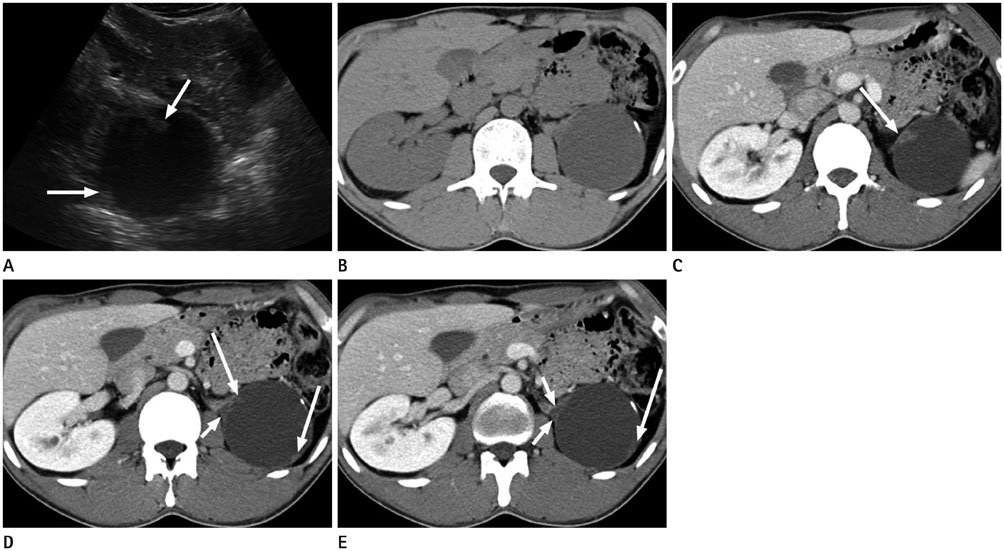
Fig. 1 33-year-old man with a left retroperitoneal mass. A. Ultrasonogram of the left intercostal space shows a cystic mass with multiple, discrete nodularites (arrows) in the left renal fossa. However, the left kidney was not clearly visible. B. Unenhanced CT image shows a cystic mass with a linear calcification. C-E. Contrast-enhanced CT images obtained during the nephrographic phase show several focal nodular enhancements (long arrows) in the cyst wall. Note the triangular, regressed, multicystic dysplastic kidney (short arrows) between the adrenal gland and the cystic mass. The normal parenchyma of the left kidney is not visible.
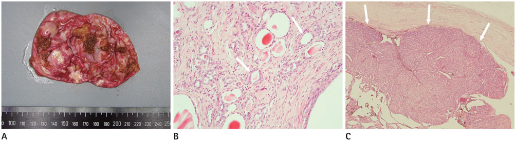
Fig. 2 Histopathologic findings of a surgically resected, left retroperitoneal mass. A. The cut surface of the surgical specimen shows tan papillary tumor tissue on the inner surface of the wall. B. Photomicrograph showing immature, tubule-like structures scattered in the fibrous cystic wall (arrows), which is a dysplastic kidney (hematoxylin and eosin stain, × 100). C. Photomicrograph showing low-grade renal cell carcinoma with papillary arrangement (arrows) (hematoxylin and eosin stain, × 40).
Reference
-
1. Matsell DG, Bennett T, Goodyer P, Goodyer C, Han VK. The pathogenesis of multicystic dysplastic kidney disease: insights from the study of fetal kidneys. Lab Invest. 1996; 74:883–893.2. Weinstein A, Goodman TR, Iragorri S. Simple multicystic dysplastic kidney disease: end points for subspecialty follow-up. Pediatr Nephrol. 2008; 23:111–116.3. Cambio AJ, Evans CP. Outcomes and quality of life issues in the pharmacological management of benign prostatic hyperplasia (BPH). Ther Clin Risk Manag. 2007; 3:181–196.4. Barrett DM, Wineland RE. Renal cell carcinoma in multicystic dysplastic kidney. Urology. 1980; 15:152–154.5. Birken G, King D, Vane D, Lloyd T. Renal cell carcinoma arising in a multicystic dysplastic kidney. J Pediatr Surg. 1985; 20:619–621.6. Rackley RR, Angermeier KW, Levin H, Pontes JE, Kay R. Renal cell carcinoma arising in a regressed multicystic dysplastic kidney. J Urol. 1994; 152(5 Pt 1):1543–1545.7. Cambio AJ, Evans CP, Kurzrock EA. Non-surgical management of multicystic dysplastic kidney. BJU Int. 2008; 101:804–808.8. Prasad SR, Humphrey PA, Catena JR, Narra VR, Srigley JR, Cortez AD, et al. Common and uncommon histologic subtypes of renal cell carcinoma: imaging spectrum with pathologic correlation. Radiographics. 2006; 26:1795–1806. discussion 1806-1810
