Korean J Obstet Gynecol.
2011 May;54(5):269-272. 10.5468/KJOG.2011.54.5.269.
A case of immature sacrococcygeal teratoma diagnosed by prenatal ultrasonography
- Affiliations
-
- 1Department of Obstetrics and Gynecology, Soonchunhyang University Cheonan Hospital, Soonchunhyang University College of Medicine, Cheonan, Korea. drsook@schmc.ac.kr
- 2Department of Pathology, Soonchunhyang University Cheonan Hospital, Soonchunhyang University College of Medicine, Cheonan, Korea.
- KMID: 2078024
- DOI: http://doi.org/10.5468/KJOG.2011.54.5.269
Abstract
- Sacrococcygeal teratoma (SCT) is a rare subset of germ cell neoplasm and occurs in approximately 1 in 35,000 live births. Most SCTs are benign, but about 20% are malignant. They originate from totipotent cells from Hansen's node or primitive germ cells, but the exact etiology remains uncertain. Antenatal diagnosis of SCT can be made by ultrasound. The fetus with SCT remains at high risk for perinatal complications and death. Perinatal mortality and morbidity are most strongly related to high-output cardiac failure because of arteriovenous shunting within the tumor, subsequent fetal hydrops, polyhydramnios, and preterm delivery. Recently we have experienced a case of immature SCT with hydrops and polyhydrmnios diagnosed by prenatal ultrasonography at 21+5 weeks, resulted in stillbirth. We describe this case with a brief review of the literature.
MeSH Terms
Figure
-
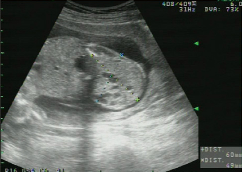
Fig. 1 Transabdominal sonogram showed the 60 × 49 mm sized sacrococcygeal teratoma.
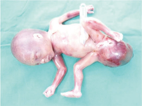
Fig. 2 Anterior gross findings of 21+5 weeks sized fetus with sacrococcygeal teratoma.
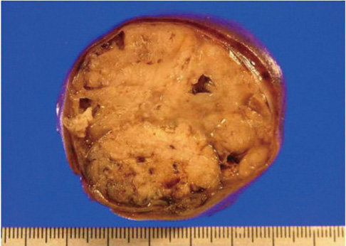
Fig. 3 Cut section showing solid, glistening mass of solid tissue and cystic spaces.
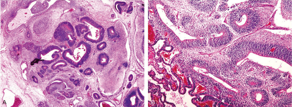
Fig. 4 (A) Highly cellular neuroglia forming neuroepithelial tubules. Cystic spaces were lined by stratified squamous epithelium (H&E, ×100). (B) Neuroepithelial tubules lined crowded cells showing loss of polarity (H&E, ×200).
Reference
-
1. Su CF, Cheung LH, Chen GD, Shih YT, Lee MS, WuH TT. Sacrococcygeal immature teratoma in a twin pregnancy. Taiwan J Obstet Gynecol. 2005. 44:196–199.2. Hirose S, Farmer DL. Fetal surgery for sacrococcygeal teratoma. Clin Perinatol. 2003. 30:493–506.3. Altman RP, Randolph JG, Lilly JR. Sacrococcygeal teratoma: American Academy of Pediatrics Surgical Section Survey-1973. J Pediatr Surg. 1974. 9:389–398.4. Flake AW. Fetal sacrococcygeal teratoma. Semin Pediatr Surg. 1993. 2:113–120.5. Sheth S, Nussbaum AR, Sanders RC, Hamper UM, Davidson AJ. Prenatal diagnosis of sacrococcygeal teratoma: sonographic-pathologic correlation. Radiology. 1988. 169:131–136.6. Holterman AX, Filiatrault D, Lallier M, Youssef S. The natural history of sacrococcygeal teratomas diagnosed through routine obstetric sonogram: a single institution experience. J Pediatr Surg. 1998. 33:899–903.7. Axt-Fliedner R, Qush H, Hendrik HJ, Ertan K, Lindinger A, Mäusle R, et al. Prenatal diagnosis of cerebral lesions and multiple intracardiac rhabdomyomas in a fetus with tuberous sclerosis. J Ultrasound Med. 2001. 20:63–67.8. Srivastava A, Jaiswal AK, Jain K, Behari S. Sacrococcygeal teratoma. J Pediatr Neurosci. 2010. 5:30–31.9. Roman AS, Monteagudo A, Timor-Tritsch I, Rebarber A. First-trimester diagnosis of sacrococcygeal teratoma: the role of three-dimensional ultrasound. Ultrasound Obstet Gynecol. 2004. 23:612–614.10. Sun DJ, Lee JN, Long CY, Tsai EM. Early diagnosis of fetal sacrococcygeal teratoma: a case report. Kaohsiung J Med Sci. 2003. 19:313–316.11. Adzick NS, Crombleholme TM, Morgan MA, Quinn TM. A rapidly growing fetal teratoma. Lancet. 1997. 349:538.12. Hedrick HL, Flake AW, Crombleholme TM, Howell LJ, Johnson MP, Wilson RD, et al. Sacrococcygeal teratoma: prenatal assessment, fetal intervention, and outcome. J Pediatr Surg. 2004. 39:430–438.13. Sherowsky RC, Williams CH, Nichols VB, Singh KB. Prenatal ultrasonographic diagnosis of a sacrococcygeal teratoma in twin pregnancy. J Ultrasound Med. 1985. 4:159–161.14. Brace V, Grant SR, Brackley KJ, Kilby MD, Whittle MJ. Prenatal diagnosis and outcome in sacrococcygeal teratomas: a review of cases between 1992 and 1998. Prenat Diagn. 2000. 20:51–55.15. Kirkinen P, Partanen K, Merikanto J, Ryynänen M, Haring P, Heinonen K. Ultrasonic and magnetic resonance imaging of fetal sacrococcygeal teratoma. Acta Obstet Gynecol Scand. 1997. 76:917–922.
- Full Text Links
-
- Actions
-
Cited
- CITED
-
- Close
- Share
-
- Similar articles
-
- A Case of Sacrococcygeal Teratoma Diagnosed by Ultrasonography and MRI at 28 Weeks of Gestation
- A Case of Sacrococcygeal Teratoma which was Antenatal Diagnosed by Ultrasonography at 35 weeks` Gestatio
- A case of Sacrococcygeal Teratoma which was Antental Diagnosed by Ultrasonography
- A case of congenital retroperitoneal immature teratoma
- A Case of Fetal Faciocervical Immature Teratoma
