Detection of chemosensitivity using K18-Asp(396) (M30) antibody in HeLa and OVCAR-3 cell lines treated with anticancer agents
- Affiliations
-
- 1Saem Hospital, Anyang-si, Gyeonggi-do, Korea.
- 2Clinical Research Laboratory, St. Mary's Hospital, The Catholic University of Korea, Seoul, Korea.
- 3Department of Obstetrics and Gynecology, The Catholic University of Korea, Seoul, Korea. nowonhkt@catholic.ac.kr
- KMID: 2077959
- DOI: http://doi.org/10.5468/kjog.2010.53.1.43
Abstract
OBJECTIVE
The aim of this study was to detect the levels of M30-antigens as a biomarker of apoptosis in cells and their culture media after treatments with anticancer drugs as a preclinical study.
METHODS
After HeLa and OVCAR-3 cells were treated respectively with paclitaxel, cisplatin, and camptothecin, the harvested cells were stained sequentially with M30 monoclonal antibodies and propidium iodide (PI). Afterwards, they were analyzed using a FACScan flow cytometer and observed under an immunofluorescence microscope for M30-FITC immunofluorescences. Levels of M30 antigens were also detected in their culture media using M30-Apoptosense ELISA kit.
RESULTS
The levels of M30-FITC immunofluorescences were elevated in both cell lines after each drug treatments compared with those of control cells. The levels of M30 antigens detected by ELISA in media culturing each cell line treated with each of drugs were elevated compared with those of control cells.
CONCLUSION
This study suggests that M30-antigens representing chemotherapy induced apoptosis may be a useful biomarker for predicting and monitoring the response of neoadjuvant chemotherapy in patients with gynecologic cancers.
Keyword
MeSH Terms
Figure
-

Fig. 1 DNA histograms (left) and linear diagrams (right) showing the changes of cell cycle phases and apoptotic populations (sub-G1 peaks) stained with propodium iodide (P1) and measured by FACScan flow cytometer in HeLa and OVCAR-3 cells. The blue peaks represent apoptotic populations (sub-G0G1 peaks). The first red peaks). The first red peaks represent the G0G1 phases of the cell cycles. The second red peaks represent G2M phases of cell cycles. The mid portions between two red peaks with diagonal lines represents S phases of cell cycles. (A) Untreated control cells, (B) cells treated with 1,000 nM of paclitaxel, (C) cells treated with 250 ng/mL of cisplatin, and (D) cells treated with 30 nM camptothecin. (E) and (F) The linear diagrams showing distributions of cell cycle phases and apoptotic sub-G0G1 peaks in HeLa and OVCAR-3 cells teated with 1,000 nM of paclitaxel, 250 ng/mL of cisplatin, and 30 nM of camptothecin.
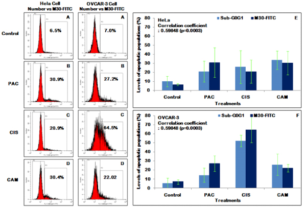
Fig. 2 The histograms are showing the levels of M30-FITC immunofluorescences (Number vs M30-FITC immunofluorescence) detected by flow cytometer in HeLa and OVCAR-3 cells treated with anti-cancer agents and the comparisons between sub-G0G1 phase fractions and positive M30-FITC immunofluorescences. The popilations in gate R2 indicate negative M30-FITC immunofluorescences and those in gate R3 indicate the apoptotic populations with positive M30-FITC immunofluorescences. (A) Untreated control cells, (B) cells treated with 1,000 nM of paclitaxel, (C) cells treated with 250 ng/mL of cisplatin, and (D) cells treated with 30 nM of camptothecin. (E) and (F) The correlation between the detected levels of sub-G0G1 fractions and M30-FITC immunofluorescences were evaluated by Spearman's Methods (correlation coefficient=0.59848, P=0.0003). There were no significant differences between them when compared by Wilcoxon Rank Sum Test.
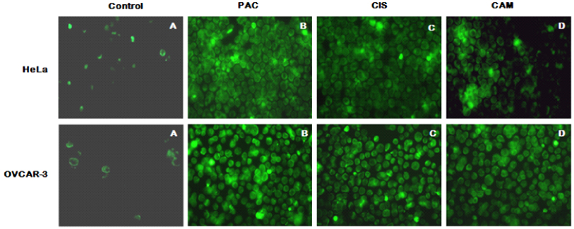
Fig. 3 The M30-FITC immunofluorescences observed under the immunofluorescence microscopy. (A) Untreated control HeLa and OVCAR-3 cells. There can be seen some positive cells with scanty intracytoplasmic M30-FITC immunofluorescences compared to the treated cells. (B~D) Cells treated with 1,000 nM of paclitaxel, 250 ug/mL of cisplatin, and 30 uM of camptothecin in order. There can be seen many cells with strong intracytoplasmic immunofluorescences in all of the treated cells, PAC: paclitaxel, CIS: cisplatin, CAM: camptothecin.
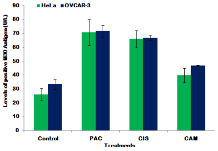
Fig. 4 The levels of M30 antigen in each cell line groups. This figure showing the comparison between the levels of CK18-Asp396 (M30) antigen detected by ELISA in the culture media of untreated controls and HeLa and OVCAR-3 cell lines treated with 1,000 nM of paclitaxel, 250 ng/mL of cisplatin, and 30 uM of camptothecin. PAC: paclitaxel, CIS: cisplatin, CAM: camptothecin.
Reference
-
1. Ayhan A, Celik H, Dursun P, Salman MC, Yuce K. Neoadjuvant chemotherapy in gynecological cancers. Eur J Gynaecol Oncol. 2006. 27:11–15.2. Richardson ME, Siemann DW. Tumor cell heterogeneity: impact on mechanisms of therapeutic drug resistance. Int J Radiat Oncol Biol Phys. 1997. 39:789–795.3. Maddika S, Ande SR, Panigrahi S, Paranjothy T, Weglarczyk K, Zuse A, et al. Cell survival, cell death and cell cycle pathways are interconnected: implications for cancer therapy. Drug Resist Updat. 2007. 10:13–29.4. Ueno T, Toi M, Linder S. Detection of epithelial cell death in the body by cytokeratin 18 measurement. Biomed Pharmacother. 2005. 59 Suppl 2:S359–S362.5. Kramer G, Erdal H, Mertens HJ, Nap M, Mauermann J, Steiner G, et al. Differentiation between cell death modes using measurements of different soluble forms of extracellular cytokeratin 18. Cancer Res. 2004. 64:1751–1756.6. Tiezzi DG, De Andrade JM, Cândido dos Reis FJ, Marana HR, Ribeiro-Silva A, Tiezzi MG, et al. Apoptosis induced by neoadjuvant chemotherapy in breast cancer. Pathology. 2006. 38:21–27.7. Ormerod MG. Investigating the relationship between the cell cycle and apoptosis using flow cytometry. J Immunol Methods. 2002. 265:73–80.8. Linder S. Cytokeratin Markers Come of Age. Tumour Biol. 2007. 28:189–195.9. Gibb RK, Taylor DD, Wan T, O'Connor DM, Doering DL, Gerçel-Taylor C. Apoptosis as a measure of chemosensitivity to cisplatin and taxol therapy in ovarian cancer cell lines. Gynecol Oncol. 1997. 65:13–22.10. Goossens JF, Hénichart JP, Dassonneville L, Facompré M, Bailly C. Relation between intracellular acidification and camptothecin- induced apoptosis in leukemia cells. Eur J Pharm Sci. 2000. 10:125–131.11. Corver WE, Cornelisse CJ, Fleuren GJ. Simultaneous measurement of two cellular antigens and DNA using fluorescein- isothiocyanate, phycoerythrin and propidium iodide on a standard FACScan. Cytometry. 1994. 15:117–128.12. Han KT, Ryu KS, Han SH, In K, Song JM, Kim JH, et al. Multiparametric flow cytometry in breast cancer cell line (MCF-7) stained with fluorescein isothiocyanate, phycoerythrin, and propidium iodide. J Korean Cancer Assoc. 1999. 31:1129–1139.13. Napolitano U, Imperato F, Mossa B, Framarino ML, Marziani R, Marzetti L. The role of neoadjuvant chemotherapy for squamous cell cervical cancer (Ib-IIIb): a long-term randomized trial. Eur J Gynaecol Oncol. 2003. 24:51–59.14. Untch M, Ditsch N, Langer E, Kurbacher C, Crohns C, Konecny G, et al. Chemosensitivity testing in gynecologic oncology--dream or reality? Recent Results Cancer Res. 2003. 161:146–158.15. Ricci MS, Zong WX. Chemotherapeutic approaches for targeting cell death pathways. Oncologist. 2006. 11:342–357.16. Leers MP, Kölgen W, Björklund V, Bergman T, Tribbick G, Persson B, et al. Immunocytochemical detection and mapping of a cytokeratin 18 neo-epitope exposed during early apoptosis. J Pathol. 1999. 187:567–572.17. Woods CM, Zhu J, McQueney PA, Bollag D, Lazarides E. Taxol-induced mitotic block triggers rapid onset of a p53-independent apoptotic pathway. Mol Med. 1995. 1:506–526.18. Wang TH, Wang HS, Soong YK. Paclitaxel- induced cell death : Where the cell cycle and apoptosis come together. Cancer. 2000. 88:2619–2628.19. Wang D, Lippard SJ. Cellular processing of platinum anticancer drugs. Nat Rev Drug Discov. 2005. 4:307–320.20. Darzynkiewicz Z, Bruno S, Del Bino G, Traganos F. The cell cycle effects of camptothecin. Ann N Y Acad Sci. 1996. 803:93–100.21. Mosesso P, Pichierri P, Franchitto A, Palitti F. Evidence that camptothecin-induced aberrations in the G2 phase of cell cycle of Chinese hamster ovary (CHO) cell lines is associated with transcription. Mutat Res. 2000. 452:189–195.22. Ueno T, Toi M, Bivén K, Bando H, Ogawa T, Linder S. Measurement of an apoptotic product in the sera of breast cancer patients. Eur J Cancer. 2003. 39:769–774.23. Ulukaya E, Yilmaztepe A, Akgoz S, Linder S, Karadag M. The levels of caspase-cleaved cytokeratin 18 are elevated in serum from patients with lung cancer and helpful to predict the survival. Lung Cancer. 2007. 56:399–404.
- Full Text Links
-
- Actions
-
Cited
- CITED
-
- Close
- Share
-
- Similar articles
-
- Measurement of apoptosis using M30 in culture media of cell lines treated with anti-cancer agents
- The Effect of Hyperthermia on Chemosensitivity of Renal Cell Carcinoma Cell Lines in vitro
- Relationship between the Expression of Apoptosis-Related Proteins and Chemosensitivity in Gastric Cancer Cell Lines
- The Expression of Multidrug Resistance-Associated Protein (MRP) and the Chemosensitivity in Gastric Cancer Cell Lines
- Flow Cytometric Detection of Apoptosis and p53 Protein in OVCAR-3 and SKOV-3 Ovarian Cancer Cell Lines
