N-acetylcysteine protects against cadmium-induced oxidative stress in rat hepatocytes
- Affiliations
-
- 1College of Veterinary Medicine, Yangzhou University, Yangzhou 225009, China. liuzongping@yzu.edu.cn
- 2College of Animal Science and Technology, Henan University of Science and Technology, Luoyang 471003, China.
- KMID: 2070234
- DOI: http://doi.org/10.4142/jvs.2014.15.4.485
Abstract
- Cadmium (Cd) is a well-known hepatotoxic environmental pollutant. We used rat hepatocytes as a model to study oxidative damage induced by Cd, effects on the antioxidant systems, and the role of N-acetylcysteine (NAC) in protecting cells against Cd toxicity. Hepatocytes were incubated for 12 and 24 h with Cd (2.5, 5, 10 microM). Results showed that Cd can induce cytotoxicity: 10 microM resulted in 36.2% mortality after 12 h and 47.8% after 24 h. Lactate dehydrogenase, aspartate aminotransferase, and alanine aminotransferase activities increased. Additionally, reactive oxygen species (ROS) generation increased in Cd-treated hepatocytes along with malondialdehyde levels. Glutathione concentrations significantly decreased after treatment with Cd for 12 h but increased after 24 h of Cd exposure. In contrast, glutathione peroxidase activity significantly increased after treatment with Cd for 12 h but decreased after 24 h. superoxide dismutase and catalase activities increased at 12 h and 24 h. glutathione S-transferase and glutathione reductase activities decreased, but not significantly. Rat hepatocytes incubated with NAC and Cd simultaneously had significantly increased viability and decreased Cd-induced ROS generation. Our results suggested that Cd induces ROS generation that leads to oxidative stress. Moreover, NAC protects rat hepatocytes from cytotoxicity associated with Cd.
Keyword
MeSH Terms
-
Acetylcysteine/*metabolism
Animals
Antioxidants/*metabolism
Cadmium/*toxicity
Cell Survival/drug effects
Cells, Cultured
Environmental Pollutants/*toxicity
Hepatocytes/drug effects/metabolism
*Oxidative Stress
Rats
Rats, Sprague-Dawley
Reactive Oxygen Species/*metabolism
Acetylcysteine
Antioxidants
Cadmium
Environmental Pollutants
Reactive Oxygen Species
Figure
-
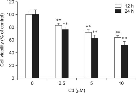
Fig. 1 Effect of cadmium (Cd) on the viability of rat hepatocytes. The cells were incubated with 0, 2.5, 5, and 10 µM Cd for 12 and 24 h. Viability was assessed by 3-(4,5-dimethylthiazol-2-yl)-2,5-diphenyl-tetrazolium bromide (MTT) colorimetric assay. Each experiment was repeated six times and data are expressed as the mean ± SD (**p < 0.01).

Fig. 2 Effects of Cd on (A) lactate dehydrogenase (LDH) activity, (B) aspartate aminotransferase (AST) activity, and (C) alanine aminotransferase (ALT) activity in rat hepatocytes. Cells were incubated with 0, 2.5, 5, and 10 µM Cd for 12 and 24 h. Each experiment was repeated six times and data are expressed as the mean ± SD (**p < 0.01).

Fig. 3 Effect of Cd on reactive oxygen species (ROS) production. Hepatocytes were treated with 5 µM of Cd for 45 min or 1.5, 6, 12, and 24 h. The cells were then stained with DCFH2-DA. Intracellular ROS levels were measured by flow cytometry as described in the Materials and Methods section. Results are expressed as a representative histogram (A, control; B, 5 µM Cd; respectively 1.5 h) and mean fluorescence obtained from the histogram statistics (C). Each bar represents the mean ± SD (n = 3). **p < 0.01 and *p < 0.05 compared to the control. DCF: dichlorofluorescein, FITC: fluorescein isothiocyanate.

Fig. 4 Effects of Cd on (A) glutathione (GSH) and (B) malondialdehyde (MDA) levels in rat hepatocytes. Cells were treated with 0, 2.5, 5, and 10 µM Cd for 12 and 24 h. Each experiment was repeated six times and data are expressed as the mean ± SD (*p < 0.05 and **p < 0.01).

Fig. 5 Effects of Cd on (A) glutathione peroxidase (GPx), (B) glutathione S-transferase (GST), (C) glutathione reductase (GR), (D) superoxide dismutase (SOD), and (E) catalase (CAT) activities. Hepatocytes were treated with 2.5, 5, and 10 µM Cd. GPx, GST, GR, SOD, and CAT activities were measured after 12 and 24 h. Each experiment was repeated six times and data are expressed as the mean ± SD (*p < 0.05 and **p < 0.01).
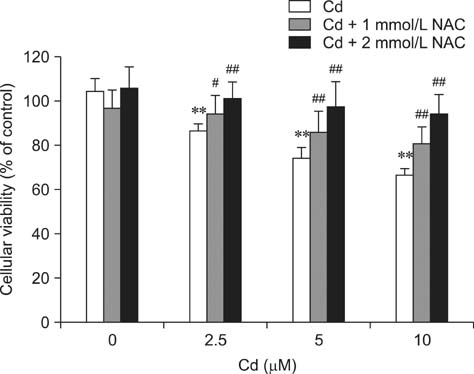
Fig. 6 Effect of N-acetylcysteine (NAC) on Cd-induced cytotoxicity in hepatocytes. Cells were incubated with NAC (1 or 2 mM) and Cd (2.5, 5, or 10 µM) simultaneously for 24 h. An MTT assay was then performed to evaluate cytotoxicity. Each experiment was repeated six times and data are expressed as the mean ± SD (**p < 0.01, #p < 0.05, and ##p < 0.01 compared to cells treated with Cd alone).

Fig. 7 Inhibitory effect of NAC on Cd-induced ROS generation (as monitored by DCF fluorescence). The cells were incubated with NAC (2 mM) and Cd (5 µM) at the same time for 1.5 h. Bars represent the mean ± SD (n = 3). **p < 0.01 and #p < 0.05 compared to cells treated with Cd alone.
Reference
-
1. Belyaeva EA, Korotkov SM. Mechanism of primary Cd2+-induced rat liver mitochondria dysfunction: discrete modes of Cd2+ action on calcium and thiol-dependent domains. Toxicol Appl Pharmacol. 2003; 192:56–68.
Article2. Bolduc JS, Denizeau F, Jumarie C. Cadmium-induced mitochondrial membrane-potential dissipation does not necessarily require cytosolic oxidative stress: studies using rhodamine-123 fluorescence unquenching. Toxicol Sci. 2004; 77:299–306.
Article3. Bonaventura J, Schroeder WA, Fang S. Human erythrocyte catalase: an improved method of isolation and a reevaluation of reported properties. Arch Biochem Biophys. 1972; 150:606–617.
Article4. Bridges CC, Zalups RK. Molecular and ionic mimicry and the transport of toxic metals. Toxicol Appl Pharmacol. 2005; 204:274–308.
Article5. Chance B, Greenstein DS, Roughton FJW. The mechanism of catalase action. I. Steady state analysis. Arch Biochem Biophys. 1952; 37:301–321.6. Chin TA, Templeton DM. Protective elevations of glutathione and metallothionein in cadmium-exposed mesangial cells. Toxicology. 1993; 77:145–156.
Article7. Dalton TP, Shertzer HG, Puga A. Regulation of gene expression by reactive oxygen. Annu Rev Pharmacol Toxicol. 1999; 39:67–101.
Article8. Ellman GL. Tissue sulfhydryl groups. Arch Biochem Biophys. 1959; 82:70–77.
Article9. Esterbauer H, Cheeseman KH. Determination of aldehydic lipid peroxidation products: malonaldehyde and 4-hydroxynonenal. Methods Enzymol. 1990; 186:407–421.10. Flohé L, Günzler WA. Assays of glutathione peroxidase. Methods Enzymol. 1984; 105:114–121.11. Funakoshi T, Ueda K, Shimada H, Kojima S. Effects of dithiocarbamates on toxicity of cadmium in rat primary hepatocyte cultures. Toxicology. 1997; 116:99–107.
Article12. Gaubin Y, Vaissade F, Croute F, Beau B, Soleilhavoup JP, Murat JC. Implication of free radicals and glutathione in the mechanism of cadmium-induced expression of stress proteins in the A549 human lung cell-line. Biochim Biophys Acta. 2000; 1495:4–13.
Article13. Habig WH, Pabst MJ, Jakoby WB. Glutathione S-transferases. The first enzymatic step in mercapturic acid formation. J Biol Chem. 1974; 249:7130–7139.14. Hatcher EL, Chen Y, Kang YJ. Cadmium resistance in A549 cells correlates with elevated glutathione content but not antioxidant enzymatic activities. Free Radic Biol Med. 1995; 19:805–812.
Article15. Hinkle PM, Kinsella PA, Osterhoudt KC. Cadmium uptake and toxicity via voltage-sensitive calcium channels. J Biol Chem. 1987; 262:16333–16337.
Article16. Hussain T, Shukla GS, Chandra SV. Effects of cadmium on superoxide dismutase and lipid peroxidation in liver and kidney of growing rats: in vivo and in vitro studies. Pharmacol Toxicol. 1987; 60:355–358.
Article17. Kakkar P, Das B, Viswanathan PN. A modified spectrophotometric assay of superoxide dismutase. Indian J Biochem Biophys. 1984; 21:130–132.18. Ketterer B. Detoxication reactions of glutathione and glutathione transferases. Xenobiotica. 1986; 16:957–973.
Article19. Kreamer BL, Staecker JL, Sawada N, Sattler GL, Hsia MT, Pitot HC. Use of a low-speed, iso-density percoll centrifugation method to increase the viability of isolated rat hepatocyte preparations. In Vitro Cell Dev Biol. 1986; 22:201–211.
Article20. Leelavinothan P, Kalist S. Beneficial effect of hesperetin on cadmium induced oxidative stress in rats: an in vivo and in vitro study. Eur Rev Med Pharmacol Sci. 2011; 15:992–1002.21. Lemarié A, Lagadic-Gossmann D, Morzadec C, Allain N, Fardel O, Vernhet L. Cadmium induces caspase-independent apoptosis in liver Hep3B cells: role for calcium in signaling oxidative stress-related impairment of mitochondria and relocation of endonuclease G and apoptosis-inducing factor. Free Radic Biol Med. 2004; 36:1517–1531.
Article22. Lin WC, Liao YC, Liau MC, Lii CK, Sheen LY. Inhibitory effect of CDA-II, a urinary preparation, on aflatoxin B1-induced oxidative stress and DNA damage in primary cultured rat hepatocytes. Food Chem Toxicol. 2006; 44:546–551.
Article23. Liu T, He W, Yan C, Qi Y, Zhang Y. Roles of reactive oxygen species and mitochondria in cadmium-induced injury of liver cells. Toxicol Ind Health. 2011; 27:249–256.
Article24. López E, Arce C, Oset-Gasque MJ, Cañadas S, González MP. Cadmium induces reactive oxygen species generation and lipid peroxidation in cortical neurons in culture. Free Radic Biol Med. 2006; 40:940–951.
Article25. Méplan C, Mann K, Hainaut P. Cadmium induces conformational modifications of wild-type p53 and suppresses p53 response to DNA damage in cultured cells. J Biol Chem. 1999; 274:31663–31670.
Article26. Mosmann T. Rapid colorimetric assay for cellular growth and survival: application to proliferation and cytotoxicity assays. J Immunol Methods. 1983; 65:55–63.
Article27. Müller L. Consequences of cadmium toxicity in rat hepatocytes: mitochondrial dysfunction and lipid peroxidation. Toxicology. 1986; 40:285–295.
Article28. Murugavel P, Pari L. Effects of diallyl tetrasulfide on cadmium-induced oxidative damage in the liver of rats. Hum Exp Toxicol. 2007; 26:527–534.
Article29. Nemmiche S, Chabane-Sari D, Guiraud P. Role of α-tocopherol in cadmium-induced oxidative stress in Wistar rat's blood, liver and brain. Chem Biol Interact. 2007; 170:221–230.
Article30. Newairy AA, El-Sharaky AS, Badreldeen MM, Eweda SM, Sheweita SA. The hepatoprotective effects of selenium against cadmium toxicity in rats. Toxicology. 2007; 242:23–30.
Article31. Nzengue Y, Steiman R, Garrel C, Lefèbvre E, Guiraud P. Oxidative stress and DNA damage induced by cadmium in the human keratinocyte HaCaT cell line: role of glutathione in the resistance to cadmium. Toxicology. 2008; 243:193–206.
Article32. Odewumi CO, Badisa VLD, Le UT, Latinwo LM, Ikediobi CO, Badisa RB, Darling-Reed SF. Protective effects of N-acetylcysteine against cadmium-induced damage in cultured rat normal liver cells. Int J Mol Med. 2011; 27:243–248.
Article33. Poliandri AHB, Cabilla JP, Velardez MO, Bodo CCA, Duvilanski BH. Cadmium induces apoptosis in anterior pituitary cells that can be reversed by treatment with antioxidants. Toxicol Appl Pharmacol. 2003; 190:17–24.
Article34. Prabu SM, Muthumani M, Shagirtha K. Quercetin potentially attenuates cadmium induced oxidative stress mediated cardiotoxicity and dyslipidemia in rats. Eur Rev Med Pharmacol Sci. 2013; 17:582–595.35. Renugadevi J, Prabu SM. Quercetin protects against oxidative stress-related renal dysfunction by cadmium in rats. Exp Toxicol Pathol. 2010; 62:471–481.
Article36. Shih CM, Ko WC, Wu JS, Wei YH, Wang LF, Chang EE, Lo TY, Cheng HH, Chen CT. Mediating of caspase-independent apoptosis by cadmium through the mitochondria-ROS pathway in MRC-5 fibroblasts. J Cell Biochem. 2004; 91:384–397.
Article37. Sinha M, Manna P, Sil PC. Taurine, a conditionally essential amino acid, ameliorates arsenic-induced cytotoxicity in murine hepatocytes. Toxicol In Vitro. 2007; 21:1419–1428.
Article38. Smith IK, Vierheller TL, Thorne CA. Assay of glutathione reductase in crude tissue homogenates using 5,5'-dithiobis(2-nitrobenzoic acid). Anal Biochem. 1988; 175:408–413.
Article39. Wispriyono B, Matsuoka M, Igisu H, Matsuno K. Protection from cadmium cytotoxicity by N-acetylcysteine in LLC-PK1 cells. J Pharmacol Exp Ther. 1998; 287:344–351.
- Full Text Links
-
- Actions
-
Cited
- CITED
-
- Close
- Share
-
- Similar articles
-
- Resveratrol Protects HepG2 and Chang Liver Cells from Oxidative Stress
- Induction of Angiogenic Cytokines in Cultured RPE by Oxidative Stress
- Light Electron Microscopic Study in Rat Livers Following Cadmium Chloride Administration
- Effect of Oxidative Stress on the Expression of Gelatinases in Cultured RPE
- Hypoxia Activates Toll-like Receptor 4 Signaling in Primary Mouse Hepatocytes Through the Receptor Clustering within Lipid Rafts
