The Use of Magnetic Resonance Imaging in Predicting the Clinical Outcome of Spinal Arteriovenous Fistula
- Affiliations
-
- 1Department of Neurosurgery, Yonsei University College of Medicine, Seoul, Korea. knkim@yuhs.ac
- 2Spine and Spinal Cord Research Institute, Yonsei University College of Medicine, Seoul, Korea.
- 3Department of Neurosurgery, Kangwon National University College of Medicine, Chuncheon, Korea.
- KMID: 2070016
- DOI: http://doi.org/10.3349/ymj.2015.56.2.397
Abstract
- PURPOSE
Magnetic resonance imaging (MRI) has been used to screen and follow-up spinal dural arteriovenous fistulae (SDAVF). The purpose of this study was to evaluate the association between MRI findings and neurologic function in SDAVF. This study also investigated clinical features and treatment results of SDAVF.
MATERIALS AND METHODS
A total of 15 consecutive patients who underwent embolization or surgery for SDAVF were included. We treated seven (60%) patients with embolization and six (40%) with surgery. We analysed clinical features, MRI findings, treatment results, and neurologic function. Neurologic function was measured by the Aminoff-Logue disability scale (ALS).
RESULTS
Patients with longer levels of intramedullary high signal intensity in preoperative T2-weighted images (T2WI) exhibited worse pre- and postoperative ALS scores (r=0.557, p=0.031; r=0.530, p=0.042, Pearson correlation). Preoperative ALS score was significantly correlated with postoperative ALS score (r=0.908, p=0.000, Pearson correlation). The number of levels showing intramedullary high signal intensity in T2WI decreased significantly postoperatively (5.2+/-3.1 vs. 1.0+/-1.4, p=0.001, Wilcoxon ranked test).
CONCLUSION
The number of involved levels of high signal intensity in preoperative T2WI is useful for predicting pre- and postoperative neurologic function in SDAVF.
MeSH Terms
-
Adult
Aged
Angiography
Arteriovenous Fistula/*pathology/radiography/*surgery
Central Nervous System Vascular Malformations/*pathology/radiography/*surgery
Embolization, Therapeutic/*methods
Female
Humans
*Magnetic Resonance Imaging
Male
Middle Aged
Postoperative Period
Predictive Value of Tests
Prognosis
Retrospective Studies
Severity of Illness Index
Spinal Cord/abnormalities/*blood supply/pathology/surgery
Treatment Outcome
Figure
-

Fig. 1 Illustrative case (Case 5). A 49-year-old male presented with progressive motor weakness for 18 months. (A) Preoperative T2-weighted MRI showed spinal cord oedema and venous engorgement. (B) Preoperative angiography. The fistula is located on the dorsal surface of the spinal cord, with a large arterialized vein emanating from the nerve root sleeve. (C) Postoperative MRI shows complete resolution of spinal cord oedema and venous engorgement. (D) Postoperative angiography. The proximal draining vein and fistula are embolized. Postoperative DSA image (anteroposterior view) demonstrating obliteration of the fistula.
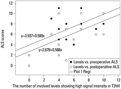
Fig. 2 The number of involved levels showing high signal intensity in T2WI vs. pre- and postoperative Aminoff-Logue disability scale (ALS) scores. T2WI, T2-weighted images.

Fig. 3 Pre- and postoperative Aminoff-Logue disability scale (ALS) scores.
Reference
-
1. da Costa L, Dehdashti AR, terBrugge KG. Spinal cord vascular shunts: spinal cord vascular malformations and dural arteriovenous fistulas. Neurosurg Focus. 2009; 26:E6.
Article2. Koch C, Kucinski T, Eckert B, Röther J, Zeumer H. [Spinal dural arteriovenous fistula: clinical and radiological findings in 54 patients]. Rofo. 2003; 175:1071–1078.3. Wakao N, Imagama S, Ito Z, Ando K, Hirano K, Tauchi R, et al. Clinical outcome of treatments for spinal dural arteriovenous fistulas: results of multivariate analysis and review of the literature. Spine (Phila Pa 1976). 2012; 37:482–488.4. Dehdashti AR, Da Costa LB, terBrugge KG, Willinsky RA, Tymianski M, Wallace MC. Overview of the current role of endovascular and surgical treatment in spinal dural arteriovenous fistulas. Neurosurg Focus. 2009; 26:E8.
Article5. Horikoshi T, Hida K, Iwasaki Y, Abe H, Akino M. Chronological changes in MRI findings of spinal dural arteriovenous fistula. Surg Neurol. 2000; 53:243–249.
Article6. Luetmer PH, Lane JI, Gilbertson JR, Bernstein MA, Huston J 3rd, Atkinson JL. Preangiographic evaluation of spinal dural arteriovenous fistulas with elliptic centric contrast-enhanced MR Angiography and effect on radiation dose and volume of iodinated contrast material. AJNR Am J Neuroradiol. 2005; 26:711–718.7. Narvid J, Hetts SW, Larsen D, Neuhaus J, Singh TP, McSwain H, et al. Spinal dural arteriovenous fistulae: clinical features and long-term results. Neurosurgery. 2008; 62:159–166.8. Steinmetz MP, Chow MM, Krishnaney AA, Andrews-Hinders D, Benzel EC, Masaryk TJ, et al. Outcome after the treatment of spinal dural arteriovenous fistulae: a contemporary single-institution series and meta-analysis. Neurosurgery. 2004; 55:77–87.
Article9. Kohno M, Takahashi H, H S, Sasaki T, Ishijima B. Functional prognosis after treatment of spinal dural arteriovenous fistulas. J Clin Neurosci. 1998; 5 Suppl:12–15.
Article10. Terwey B, Becker H, Thron AK, Vahldiek G. Gadolinium-DTPA enhanced MR imaging of spinal dural arteriovenous fistulas. J Comput Assist Tomogr. 1989; 13:30–37.
Article11. Willinsky RA, terBrugge K, Montanera W, Mikulis D, Wallace MC. Posttreatment MR findings in spinal dural arteriovenous malformations. AJNR Am J Neuroradiol. 1995; 16:2063–2071.12. Aminoff MJ, Barnard RO, Logue V. The pathophysiology of spinal vascular malformations. J Neurol Sci. 1974; 23:255–263.
Article13. Hassler W, Thron A, Grote EH. Hemodynamics of spinal dural arteriovenous fistulas. An intraoperative study. J Neurosurg. 1989; 70:360–370.14. Kendall BE, Logue V. Spinal epidural angiomatous malformations draining into intrathecal veins. Neuroradiology. 1977; 13:181–189.
Article15. Isu T, Iwasaki Y, Akino M, Koyanagi I, Abe H. Magnetic resonance imaging in cases of spinal dural arteriovenous malformation. Neurosurgery. 1989; 24:919–923.
Article16. Gokhale S, Khan SA, McDonagh DL, Britz G. Comparison of surgical and endovascular approach in management of spinal dural arteriovenous fistulas: a single center experience of 27 patients. Surg Neurol Int. 2014; 5:7.
Article17. Fisher CG, Noonan VK, Smith DE, Wing PC, Dvorak MF, Kwon BK. Motor recovery, functional status, and health-related quality of life in patients with complete spinal cord injuries. Spine (Phila Pa 1976). 2005; 30:2200–2207.
Article18. Ropper AE, Gross BA, Du R. Surgical treatment of Type I spinal dural arteriovenous fistulas. Neurosurg Focus. 2012; 32:E3.
Article19. Shin DA, Kim SH, Kim KN, Shin HC, Yoon DH. Surgical management of spinal cord haemangioblastoma. Acta Neurochir (Wien). 2008; 150:215–220.
Article20. Tacconi L, Lopez Izquierdo BC, Symon L. Outcome and prognostic factors in the surgical treatment of spinal dural arteriovenous fistulas. A long-term study. Br J Neurosurg. 1997; 11:298–305.
Article
- Full Text Links
-
- Actions
-
Cited
- CITED
-
- Close
- Share
-
- Similar articles
-
- Intracranial Dural Arteriovenous Fistula Draining into Spinal Perimedullary Veins: A Rare Cause of Myelopathy
- Myelopathy due to Spinal Dural Arteriovenous Fistula: A Case Report
- Syringomyelia Associated with Spinal Dural Arteriovenous Fistula: Clinical and Radiological Improvement after Embolization
- Spinal Dural Arteriovenous Fistula with Supply from the Lateral Sacral Artery: Case Report and Review of Literature
- Occurrence of Metachronous Intracranial Dural Arteriovenous Fistula after Embolization of Intracranial Dural Arteriovenous Fistula: A Case Report
