Role of C-Arm Cone-Beam CT in Chemoembolization for Hepatocellular Carcinoma
- Affiliations
-
- 1Department of Radiology, Seoul National University College of Medicine, Institute of Radiation Medicine, Seoul National University Medical Research Center, and Clinical Research Institute, Seoul National University Hospital, Seoul 110-744, Korea. angioint
- KMID: 2069989
- DOI: http://doi.org/10.3348/kjr.2015.16.1.114
Abstract
- With the advent of C-arm cone-beam computed tomography (CBCT), minimally-invasive procedures in the angiography suite made a new leap beyond the limitations of 2-dimensional (D) angiography alone. C-arm CBCT can help interventional radiologists in several ways with the treatment of hepatocellular carcinoma (HCC); visualization of small tumors and tumor-feeding arteries, identification of occult lesion and 3D configuration of tortuous hepatic arteries, assurance of completeness of chemoembolization, suggestion of presence of extrahepatic collateral arteries supplying HCCs, and prevention of nontarget embolization. With more improvements in the technology, C-arm CBCT may be essential in all kinds of interventional procedures in the near future.
MeSH Terms
Figure
-
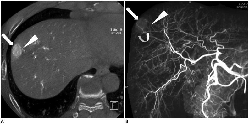
Fig. 1 60-year-old man with hepatocellular carcinoma.A. C-arm cone-beam CT shows nodular tumor (arrow) supplied by adjacent subsegmental hepatic artery (arrowhead). B. Maximum intensity projection image of C-arm cone-beam CT obtained at common hepatic artery shows small nodular tumor (arrow) supplied by subsegmental hepatic artery (arrowhead) which was noted on axial image (A). Note another subsegmental hepatic artery (curved arrow) feeding nodular tumor.
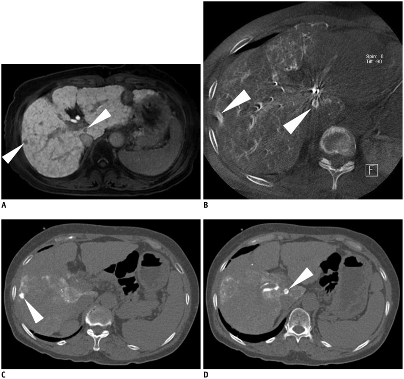
Fig. 2 2. 58-year-old woman with hepatocellular carcinoma.A. Hepatobiliary phase image of gadoxetic acid-enhanced MRI shows two small nodules of hypointensity (arrowheads). These two nodules show no enhancement on arterial phase images of MRI and on arterial phase of CT scan (not shown). B. Axial image of C-arm cone-beam CT shows enhancement of these two nodules (arrowheads). Note motion artifact of hepatic artery caused by inadequate breath-hold. C, D. Unenhanced CT scan images obtained immediately after chemoembolization show dense accumulation of iodized oil in these two nodules (arrowheads) with surrounding parenchymal accumulation of iodized oil.
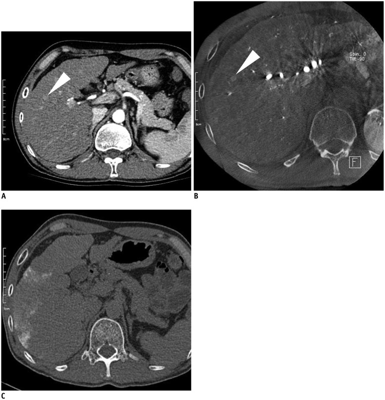
Fig. 3 42-year-old man with hepatocellular carcinoma.A. Axial CT scan shows small subtle enhancing lesion (arrowhead) in right lobe of liver. Wash-out of this nodule is equivocal on delayed image. B. Axial image of C-arm cone-beam CT shows subtle nodular enhancing lesion (arrowhead). This lesion was treated by iodized oil emulsion. C. Unenhanced CT scan obtained immediately after chemoembolization shows no nodular accumulation of iodized oil. During 2-year follow-up, this subtle nodule has persisted on follow-up CT scan without morphological change, which was thought to be benign arterioportal shunt.
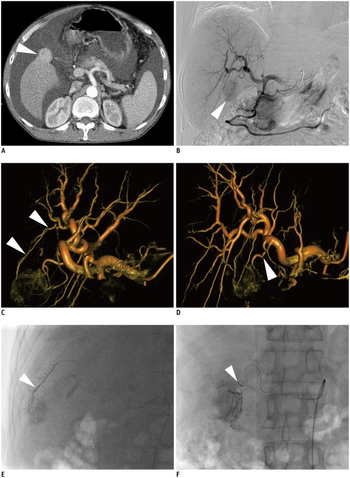
Fig. 4 47-year-old man with hepatocellular carcinoma and Child-Pugh C class disease.A. Axial CT scan shows exophytic enhancing nodule (arrowhead) in gallbladder bed. B. Celiac angiography shows tumor staining (arrowhead). C. Volume-rendering image of C-arm cone-beam CT with left anterior oblique projection of 30 degree shows tumor-feeding artery from S5 hepatic artery (arrowheads). D. Volume-rendering image of C-arm cone-beam CT with right anterior oblique projection of 20 degree and cranial oblique projection of 15 degree shows tumor-feeding artery from deep cystic artery (arrowhead). E. Spot image during chemoembolization shows tip (arrowhead) of microcatheter advanced into S5 hepatic artery. F. Spot image during chemoembolization shows tip (arrowhead) of microcatheter advanced into deep cystic artery.
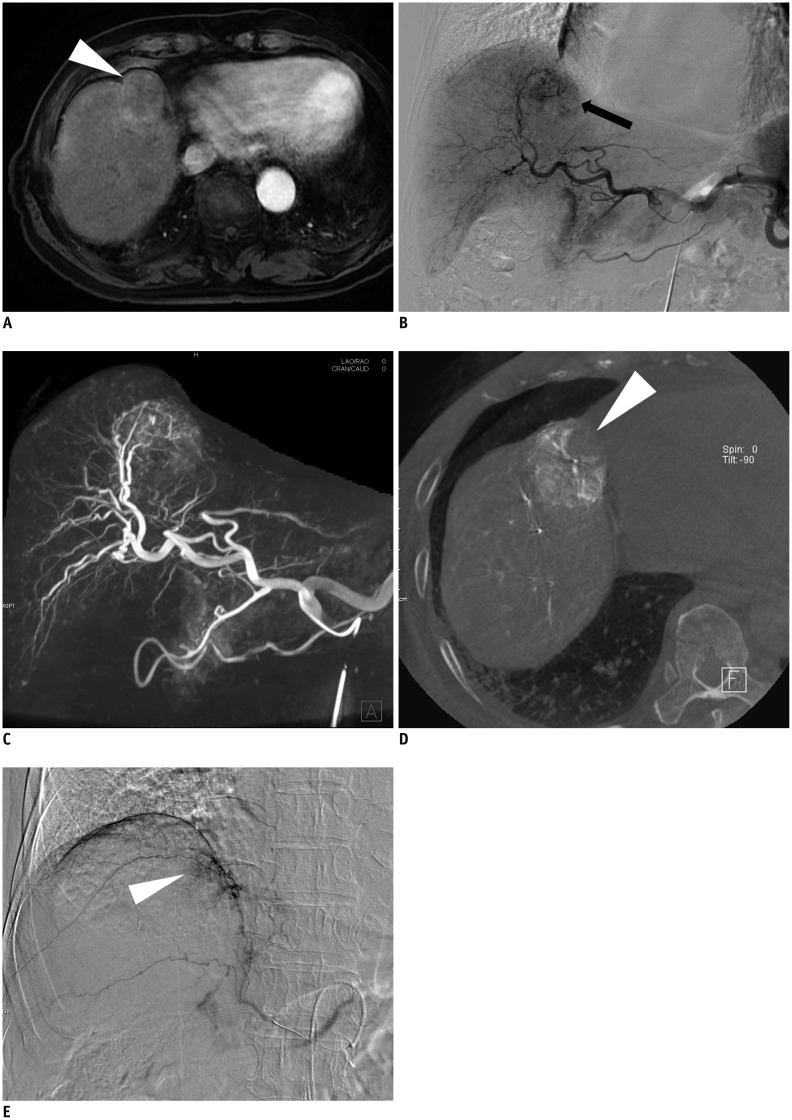
Fig. 5 78-year-old man with hepatocellular carcinoma.A. Arterial phase images of gadoxetic acid-enhanced MRI shows exophytic nodule (arrowhead) with faint enhancement. B. Celiac angiography shows hypervascular tumor staining (arrow). C. Maximum-intensity-projection image of C-arm cone-beam CT shows hypervascular tumor staining. D. Axial image of C-arm cone-beam CT shows non-enhancing part (arrowhead) of tumor which suggests presence of extrahepatic collateral artery supplying tumor. E. Angiography of right inferior phrenic artery shows tumor staining (arrowhead).
Cited by 4 articles
-
Imaging Evaluation Following 90Y Radioembolization of Liver Tumors: What Radiologists Should Know
Ijin Joo, Hyo-Cheol Kim, Gyoung Min Kim, Jin Chul Paeng
Korean J Radiol. 2018;19(2):209-222. doi: 10.3348/kjr.2018.19.2.209.Optimized Performance of FlightPlan during Chemoembolization for Hepatocellular Carcinoma: Importance of the Proportion of Segmented Tumor Area
Seung-Moon Joo, Yong Pyo Kim, Tae Jun Yum, Na Lae Eun, Dahye Lee, Kwang-Hun Lee
Korean J Radiol. 2016;17(5):771-778. doi: 10.3348/kjr.2016.17.5.771.Update on Transarterial Chemoembolization with Drug-Eluting Microspheres for Hepatocellular Carcinoma
Yasir M. Nouri, Jin Hyoung Kim, Hyun-Ki Yoon, Heung-Kyu Ko, Ji Hoon Shin, Dong Il Gwon
Korean J Radiol. 2019;20(1):34-49. doi: 10.3348/kjr.2018.0088.Detection of Recurrent/Residual Hepatocellular Carcinoma: Single-Center Retrospective Comparative Study Between Parenchymal Blood Volume Mapping Using Cone Beam CT and Multiphase Dynamic CT
Na Rae Kim, Ji Dae Kim, Hyun Young Han, Hee Jin Kim
J Korean Soc Radiol. 2018;79(2):68-76. doi: 10.3348/jksr.2018.79.2.68.
Reference
-
1. Shin SW. The current practice of transarterial chemoembolization for the treatment of hepatocellular carcinoma. Korean J Radiol. 2009; 10:425–434. PMID: 19721826.
Article2. Cheung JY, Kim Y, Shim SS, Lim SM. Combined fluoroscopy- and CT-guided transthoracic needle biopsy using a C-arm cone-beam CT system: comparison with fluoroscopy-guided biopsy. Korean J Radiol. 2011; 12:89–96. PMID: 21228944.
Article3. Georgiades CS, Hong K, Geschwind JF, Liddell R, Syed L, Kharlip J, et al. Adjunctive use of C-arm CT may eliminate technical failure in adrenal vein sampling. J Vasc Interv Radiol. 2007; 18:1102–1105. PMID: 17804771.
Article4. Kakeda S, Korogi Y, Ohnari N, Moriya J, Oda N, Nishino K, et al. Usefulness of cone-beam volume CT with flat panel detectors in conjunction with catheter angiography for transcatheter arterial embolization. J Vasc Interv Radiol. 2007; 18:1508–1516. PMID: 18057285.
Article5. Collins J, Salem R. Hepatic radioembolization complicated by gastrointestinal ulceration. Semin Intervent Radiol. 2011; 28:240–245. PMID: 22654271.
Article6. Orth RC, Wallace MJ, Kuo MD. Technology Assessment Committee of the Society of Interventional Radiology. C-arm cone-beam CT: general principles and technical considerations for use in interventional radiology. J Vasc Interv Radiol. 2008; 19:814–820. PMID: 18503894.
Article7. Tognolini A, Louie JD, Hwang GL, Hofmann LV, Sze DY, Kothary N. Utility of C-arm CT in patients with hepatocellular carcinoma undergoing transhepatic arterial chemoembolization. J Vasc Interv Radiol. 2010; 21:339–347. PMID: 20133156.
Article8. Wallace MJ, Kuo MD, Glaiberman C, Binkert CA, Orth RC, Soulez G, et al. Three-dimensional C-arm cone-beam CT: applications in the interventional suite. J Vasc Interv Radiol. 2008; 19:799–813. PMID: 18503893.
Article9. Koelblinger C, Schima W, Berger-Kulemann V, Wolf F, Plank C, Weber M, et al. C-arm CT during hepatic arteriography tumour-to-liver contrast: intraindividual comparison of three different contrast media application protocols. Eur Radiol. 2013; 23:938–942. PMID: 23138384.
Article10. Miyayama S, Yamashiro M, Okuda M, Yoshie Y, Nakashima Y, Ikeno H, et al. Detection of corona enhancement of hypervascular hepatocellular carcinoma by C-arm dual-phase cone-beam CT during hepatic arteriography. Cardiovasc Intervent Radiol. 2011; 34:81–86. PMID: 20333382.
Article11. Loffroy R, Lin M, Yenokyan G, Rao PP, Bhagat N, Noordhoek N, et al. Intraprocedural C-arm dual-phase cone-beam CT: can it be used to predict short-term response to TACE with drug-eluting beads in patients with hepatocellular carcinoma? Radiology. 2013; 266:636–648. PMID: 23143027.
Article12. Higashihara H, Osuga K, Onishi H, Nakamoto A, Tsuboyama T, Maeda N, et al. Diagnostic accuracy of C-arm CT during selective transcatheter angiography for hepatocellular carcinoma: comparison with intravenous contrast-enhanced, biphasic, dynamic MDCT. Eur Radiol. 2012; 22:872–879. PMID: 22120061.
Article13. Meyer BC, Frericks BB, Voges M, Borchert M, Martus P, Justiz J, et al. Visualization of hypervascular liver lesions During TACE: comparison of angiographic C-arm CT and MDCT. AJR Am J Roentgenol. 2008; 190:W263–W269. PMID: 18356419.
Article14. Iwazawa J, Ohue S, Hashimoto N, Abe H, Hamuro M, Mitani T. Detection of hepatocellular carcinoma: comparison of angiographic C-arm CT and MDCT. AJR Am J Roentgenol. 2010; 195:882–887. PMID: 20858813.
Article15. Miyayama S, Yamashiro M, Okuda M, Yoshie Y, Sugimori N, Igarashi S, et al. Usefulness of cone-beam computed tomography during ultraselective transcatheter arterial chemoembolization for small hepatocellular carcinomas that cannot be demonstrated on angiography. Cardiovasc Intervent Radiol. 2009; 32:255–264. PMID: 19067043.
Article16. Loffroy R, Lin M, Rao P, Bhagat N, Noordhoek N, Radaelli A, et al. Comparing the detectability of hepatocellular carcinoma by C-arm dual-phase cone-beam computed tomography during hepatic arteriography with conventional contrast-enhanced magnetic resonance imaging. Cardiovasc Intervent Radiol. 2012; 35:97–104. PMID: 21328023.
Article17. Onishi H, Kim T, Imai Y, Hori M, Nagano H, Nakaya Y, et al. Hypervascular hepatocellular carcinomas: detection with gadoxetate disodium-enhanced MR imaging and multiphasic multidetector CT. Eur Radiol. 2012; 22:845–854. PMID: 22057248.
Article18. Yu MH, Kim JH, Yoon JH, Kim HC, Chung JW, Han JK, et al. Role of C-arm CT for transcatheter arterial chemoembolization of hepatocellular carcinoma: diagnostic performance and predictive value for therapeutic response compared with gadoxetic acid-enhanced MRI. AJR Am J Roentgenol. 2013; 201:675–683. PMID: 23971463.
Article19. Meyer BC, Witschel M, Frericks BB, Voges M, Hopfenmüller W, Wolf KJ, et al. The value of combined soft-tissue and vessel visualisation before transarterial chemoembolisation of the liver using C-arm computed tomography. Eur Radiol. 2009; 19:2302–2309. PMID: 19424701.
Article20. Iwazawa J, Ohue S, Mitani T, Abe H, Hashimoto N, Hamuro M, et al. Identifying feeding arteries during TACE of hepatic tumors: comparison of C-arm CT and digital subtraction angiography. AJR Am J Roentgenol. 2009; 192:1057–1063. PMID: 19304714.
Article21. Choi WS, Kim HC, Hur S, Choi JW, Lee JH, Yu SJ, et al. Role of C-arm CT in identifying caudate arteries supplying hepatocellular carcinoma. J Vasc Interv Radiol. 2014; 25:1380–1388. PMID: 24713418.
Article22. Wang X, Shah RP, Maybody M, Brown KT, Getrajdman GI, Stevenson C, et al. Cystic artery localization with a three-dimensional angiography vessel tracking system compared with conventional two-dimensional angiography. J Vasc Interv Radiol. 2011; 22:1414–1419. PMID: 21546264.
Article23. Deschamps F, Solomon SB, Thornton RH, Rao P, Hakime A, Kuoch V, et al. Computed analysis of three-dimensional cone-beam computed tomography angiography for determination of tumor-feeding vessels during chemoembolization of liver tumor: a pilot study. Cardiovasc Intervent Radiol. 2010; 33:1235–1242.
Article24. Miyayama S, Yamashiro M, Ikuno M, Okumura K, Yoshida M. Ultraselective transcatheter arterial chemoembolization for small hepatocellular carcinoma guided by automated tumor-feeders detection software: technical success and short-term tumor response. Abdom Imaging. 2014; 39:645–656. PMID: 24549881.
Article25. Miyayama S, Yamashiro M, Hashimoto M, Hashimoto N, Ikuno M, Okumura K, et al. Identification of small hepatocellular carcinoma and tumor-feeding branches with cone-beam CT guidance technology during transcatheter arterial chemoembolization. J Vasc Interv Radiol. 2013; 24:501–508. PMID: 23452552.
Article26. Iwazawa J, Ohue S, Hashimoto N, Muramoto O, Mitani T. Clinical utility and limitations of tumor-feeder detection software for liver cancer embolization. Eur J Radiol. 2013; 82:1665–1671. PMID: 23743053.
Article27. Song SY, Chung JW, Lim HG, Park JH. Nonhepatic arteries originating from the hepatic arteries: angiographic analysis in 250 patients. J Vasc Interv Radiol. 2006; 17:461–469. PMID: 16567670.
Article28. Kim HC, Chung JW, Park JH, An S, Son KR, Seong NJ, et al. Transcatheter arterial chemoembolization for hepatocellular carcinoma: prospective assessment of the right inferior phrenic artery with C-arm CT. J Vasc Interv Radiol. 2009; 20:888–895. PMID: 19481471.
Article29. Kim HC, Chung JW, An S, Seong NJ, Jae HJ, Cho BH, et al. Left inferior phrenic artery feeding hepatocellular carcinoma: angiographic anatomy using C-arm CT. AJR Am J Roentgenol. 2009; 193:W288–W294. PMID: 19770297.
Article30. Kim HC, Chung JW, Lee IJ, An S, Seong NJ, Son KR, et al. Intercostal artery supplying hepatocellular carcinoma: demonstration of a tumor feeder by C-arm CT and multidetector row CT. Cardiovasc Intervent Radiol. 2011; 34:87–91. PMID: 20458586.
Article31. Kim HC, Chung JW, Lee W, Jae HJ, Park JH. Recognizing extrahepatic collateral vessels that supply hepatocellular carcinoma to avoid complications of transcatheter arterial chemoembolization. Radiographics. 2005; 25(Suppl 1):S25–S39. PMID: 16227494.
Article32. Iwazawa J, Ohue S, Kitayama T, Sassa S, Mitani T. C-arm CT for assessing initial failure of iodized oil accumulation in chemoembolization of hepatocellular carcinoma. AJR Am J Roentgenol. 2011; 197:W337–W342. PMID: 21785062.
Article33. Miyayama S, Yamashiro M, Hashimoto M, Hashimoto N, Ikuno M, Okumura K, et al. Comparison of local control in transcatheter arterial chemoembolization of hepatocellular carcinoma ≤6 cm with or without intraprocedural monitoring of the embolized area using cone-beam computed tomography. Cardiovasc Intervent Radiol. 2014; 37:388–395. PMID: 23775550.34. Suk Oh J, Jong Chun H, Gil Choi B, Giu Lee H. Transarterial chemoembolization with drug-eluting beads in hepatocellular carcinoma: usefulness of contrast saturation features on cone-beam computed tomography imaging for predicting short-term tumor response. J Vasc Interv Radiol. 2013; 24:483–489. PMID: 23452553.
Article35. Virmani S, Ryu RK, Sato KT, Lewandowski RJ, Kulik L, Mulcahy MF, et al. Effect of C-arm angiographic CT on transcatheter arterial chemoembolization of liver tumors. J Vasc Interv Radiol. 2007; 18:1305–1309. PMID: 17911523.
Article36. Wallace MJ, Murthy R, Kamat PP, Moore T, Rao SH, Ensor J, et al. Impact of C-arm CT on hepatic arterial interventions for hepatic malignancies. J Vasc Interv Radiol. 2007; 18:1500–1507. PMID: 18057284.
Article37. Iwazawa J, Ohue S, Hashimoto N, Muramoto O, Mitani T. Survival after C-arm CT-assisted chemoembolization of unresectable hepatocellular carcinoma. Eur J Radiol. 2012; 81:3985–3992. PMID: 22959287.
Article38. Kothary N, Abdelmaksoud MH, Tognolini A, Fahrig R, Rosenberg J, Hovsepian DM, et al. Imaging guidance with C-arm CT: prospective evaluation of its impact on patient radiation exposure during transhepatic arterial chemoembolization. J Vasc Interv Radiol. 2011; 22:1535–1543. PMID: 21875814.
Article
- Full Text Links
-
- Actions
-
Cited
- CITED
-
- Close
- Share
-
- Similar articles
-
- Conventional Chemoembolization for Hepatocellular Carcinoma: Role of Cone-Beam Computed Tomography Guidance
- Detection of Recurrent/Residual Hepatocellular Carcinoma: Single-Center Retrospective Comparative Study Between Parenchymal Blood Volume Mapping Using Cone Beam CT and Multiphase Dynamic CT
- Comparison of surgical resection versus transarterial chemoembolization with additional radiation therapy in patients with hepatocellular carcinoma with portal vein invasion
- Rapid Intra-Hepatic Dissemination of Hepatocellular Carcinoma with Pulmonary Metastases Following Combined Loco-Regional Therapy
- Incidence and Risk Factors of Acute Ischemic Cholecystitis after Transarterial Chemoembolization: Correlation with Cone Beam CT Findings
