Reversible Cerebral Vasoconstriction Syndrome and Posterior Reversible Encephalopathy Syndrome Presenting with Deep Intracerebral Hemorrhage in Young Women
- Affiliations
-
- 1Department of Neurosurgery, Samsung Medical Center, Sungkyunkwan University School of Medicine, Seoul, Korea. yeonjay@naver.com
- KMID: 2069250
- DOI: http://doi.org/10.7461/jcen.2015.17.3.239
Abstract
- Reversible cerebral vasoconstriction syndrome (RCVS) is a group of syndromes characterized by reversible segmental constriction of cerebral arteries. Posterior reversible encephalopathy syndrome (PRES) is another clinical-radiologic syndrome characterized by reversible, posterior-predominant brain edema. Although the exact causes of these reversible syndromes are poorly understood, these entities may share some common pathophysiologic elements leading to hemorrhagic strokes and rarely, deep intracerebral hemorrhage (ICH). Recent studies have suggested that endothelial dysfunction is a common pathophysiologic factor associated with these syndromes. We report on two young female patients who presented with deep ICH and were later diagnosed as RCVS and PRES. Both patients suffered from vasoconstriction and delayed ischemic stroke. Early detection of distinguishing clinical-radiologic features associated with these reversible syndromes and removing triggers would facilitate successful treatment with no complications.
Keyword
MeSH Terms
Figure
-
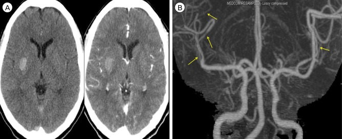
Fig. 1 (A) Brain CT shows the isolated deep ICH in the right basal ganglia. (B) Brain CTA shows multifocal narrowing of distal MCA branches (arrows). CT = computed tomography; ICH = intracerebral hemorrhage; CTA = CT angiography; MCA = middle cerebral arteries.
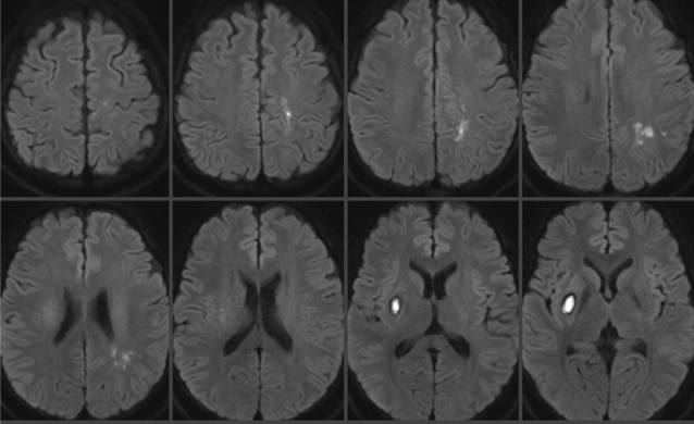
Fig. 2 MRI shows multifocal border zone infarctions. MRI = magnetic resonance imaging.
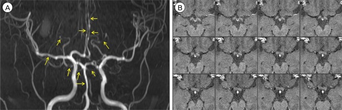
Fig. 3 (A) MRA shows the progression of multifocal vasoconstriction toward proximal cerebral arteries. (B) There is no wall enhancement of affected vessels on high resolution MR wall imaging. MRA = MR angiography; MR = magnetic resonance.

Fig. 4 Digital subtraction angiography demonstrates improving but residual vasoconstriction (arrows).
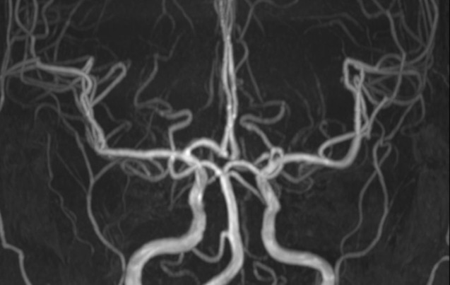
Fig. 5 Complete resolution of vasoconstriction is shown on 6-month follow-up MRA. MRA = MR angiography.
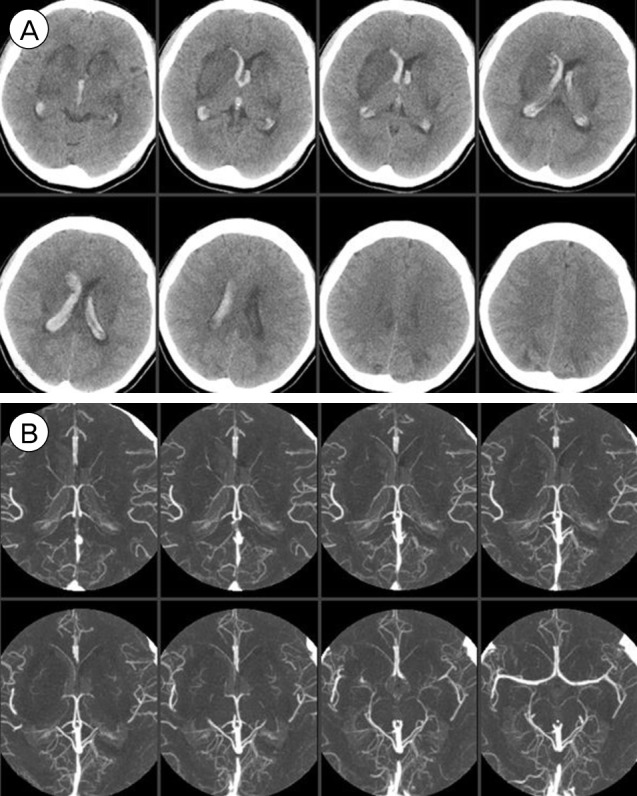
Fig. 6 (A) Brain CT shows deep ICH and associated IVH. Note the symmetrically scattered low density lesions in the bilateral basal ganglia and parietal lobes. (B) There is no vascular abnormality. CT = computed tomography; ICH = intracerebral hemorrhage; IVH = intraventricular hemorrhage.
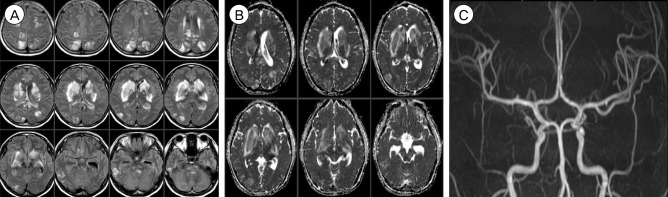
Fig. 7 (A, B) T2 Fluid Attenuated Inversion Recovery (FLAIR) images (A) and apparent diffusion coefficient images (B) suggest vasogenic edema. (C) MRA is normal. MRA = MR angiography.
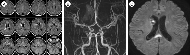
Fig. 8 (A) Follow-up MRI shows improved vasogenic edema. (B, C) Newly appeared multifocal stenoses are seen (B) with a new infarction (C). MRI = magnetic resonance imaging.
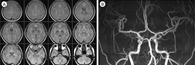
Fig. 9 Complete resolution of vasogenic edema and multifocal stenoses is shown on 3-month follow-up MRI (A) and MRA (B). MRI = magnetic resonance imaging; MRA = MR angiography.
Reference
-
1. Bartynski WS. Posterior reversible encephalopathy syndrome, part 1: fundamental imaging and clinical features. AJNR Am J Neuroradiol. 2008; 6. 29(6):1036–1042. PMID: 18356474.
Article2. Bartynski WS. Posterior reversible encephalopathy syndrome, part 2: controversies surrounding pathophysiology of vasogenic edema. AJNR Am J Neuroradiol. 2008; 6. 29(6):1043–1049. PMID: 18403560.
Article3. Bartynski WS, Boardman JF. Catheter angiography, MR angiography, and MR perfusion in posterior reversible encephalopathy syndrome. AJNR Am J Neuroradiol. 2008; 3. 29(3):447–455. PMID: 18079186.
Article4. Brandes RP. Endothelial dysfunction and hypertension. Hypertension. 2014; 11. 64(5):924–928. PMID: 25156167.
Article5. Calabrese LH, Dodick DW, Schwedt TJ, Singhal AB. Narrative review: reversible cerebral vasoconstriction syndromes. Ann Intern Med. 2007; 1. 146(1):34–44. PMID: 17200220.
Article6. Call GK, Fleming MC, Sealfon S, Levine H, Kistler JP, Fisher CM. Reversible cerebral segmental vasoconstriction. Stroke. 1988; 9. 19(9):1159–1170. PMID: 3046073.
Article7. Chen SP, Fuh JL, Wang SJ, Chang FC, Lirng JF, Fang YC, et al. Magnetic resonance angiography in reversible cerebral vasoconstriction syndromes. Ann Neurol. 2010; 5. 67(5):648–656. PMID: 20437562.
Article8. Chen SP, Fuh JL, Wang SJ, Tsai SJ, Hong CJ, Yang AC. Brain-derived neurotrophic factor gene Val66Met polymorphism modulates reversible cerebral vasoconstriction syndromes. PLoS One. 2011; 3. 6(3):e18024. PMID: 21437208.
Article9. Chen SP, Wang YF, Huang PH, Chi CW, Fuh JL, Wang SJ. Reduced circulating endothelial progenitor cells in reversible cerebral vasoconstriction syndrome. J Headache Pain. 2014; 12. 15:82. PMID: 25466718.
Article10. Dou YH, Fuh JL, Chen SP, Wang SJ. Reversible cerebral vasoconstriction syndrome after blood transfusion. Headache. 2014; 4. 54(4):736–744. PMID: 24628283.
Article11. Ducros A. Reversible cerebral vasoconstriction syndrome. Lancet Neurol. 2012; 10. 11(10):906–917. PMID: 22995694.
Article12. Ducros A, Boukobza M, Porcher R, Sarov M, Valade D, Bousser MG. The clinical and radiological spectrum of reversible cerebral vasoconstriction syndrome. A prospective series of 67 patients. Brain. 2007; 12. 130(Pt 12):3091–3101. PMID: 18025032.
Article13. Ducros A, Fiedler U, Porcher R, Boukobza M, Stapf C, Bousser MG. Hemorrhagic manifestations of reversible cerebral vasoconstriction syndrome: frequency, features, and risk factors. Stroke. 2010; 11. 41(11):2505–2511. PMID: 20884871.14. Hefzy HM, Bartynski WS, Boardman JF, Lacomis D. Hemorrhage in posterior reversible encephalopathy syndrome (PRES): Imaging and clinical features. AJNR Am J Neuroradiol. 2009; 8. 30(7):1371–1379. PMID: 19386731.15. Hinchey J, Chaves C, Appignani B, Breen J, Pao L, Wang A, et al. A reversible posterior leukoencephalopathy syndrome. N Engl J Med. 1996; 2. 334(8):494–500. PMID: 8559202.
Article16. Marder CP, Donohue MM, Weinstein JR, Fink KR. Multimodal imaging of reversible cerebral vasoconstriction syndrome: a series of 6 cases. AJNR Am J Neuroradiol. 2012; 8. 33(7):1403–1410. PMID: 22422190.
Article17. Marra A, Vargas M, Striano P, Del Guercio L, Buonanno P, Servillo G. Posterior reversible encephalopathy syndrome: the endothelial hypotheses. Med Hypotheses. 2014; 5. 82(5):619–622. PMID: 24613735.
Article
- Full Text Links
-
- Actions
-
Cited
- CITED
-
- Close
- Share
-
- Similar articles
-
- Reversible Cerebral Vasoconstriction Syndrome Combined with Posterior Encephalopathy Syndrome, and Transient Splenial Lesion after Delivery
- Progressive Manifestations of Reversible Cerebral Vasoconstriction Syndrome Presenting with Subarachnoid Hemorrhage, Intracerebral Hemorrhage, and Cerebral Infarction
- Posterior reversible encephalopathy syndrome and reversible cerebral vasoconstriction syndrome associated with acute exacerbation of chronic obstructive pulmonary disease
- A Case Report Reversible Cerebral Vasoconstriction Syndrome with Thunderclap Headache During Swimming
- Posterior Reversible Encephalopathy Syndrome in a Patient with Intoxication of Arisaema amurense
