Alveolar bone thickness around maxillary central incisors of different inclination assessed with cone-beam computed tomography
- Affiliations
-
- 1Department of Orthodontics, School of Stomatology, China Medical University, Shenyang, China. tianyulou@sina.com
- 2Department of Orthodontics, Shenyang Stomatology Hospital, Shenyang, China.
- KMID: 2068519
- DOI: http://doi.org/10.4041/kjod.2015.45.5.245
Abstract
OBJECTIVE
To assess the labial and lingual alveolar bone thickness in adults with maxillary central incisors of different inclination by cone-beam computed tomography (CBCT).
METHODS
Ninety maxillary central incisors from 45 patients were divided into three groups based on the maxillary central incisors to palatal plane angle; lingual-inclined, normal, and labial-inclined. Reformatted CBCT images were used to measure the labial and lingual alveolar bone thickness (ABT) at intervals corresponding to every 1/10 of the root length. The sum of labial ABT and lingual ABT at the level of the root apex was used to calculate the total ABT (TABT). The number of teeth exhibiting alveolar fenestration and dehiscence in each group was also tallied. One-way analysis of variance and Tukey's honestly significant difference test were applied for statistical analysis.
RESULTS
The labial ABT and TABT values at the root apex in the lingual-inclined group were significantly lower than in the other groups (p < 0.05). Lingual and labial ABT values were very low at the cervical level in the lingual-inclined and normal groups. There was a higher prevalence of alveolar fenestration in the lingual-inclined group.
CONCLUSIONS
Lingual-inclined maxillary central incisors have less bone support at the level of the root apex and a greater frequency of alveolar bone defects than normal maxillary central incisors. The bone plate at the marginal level is also very thin.
Keyword
Figure
-
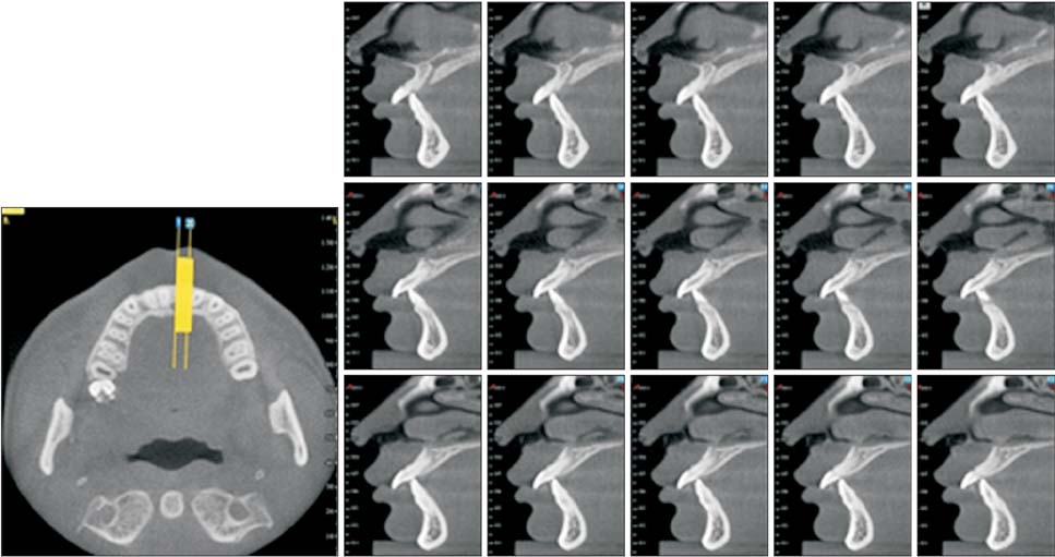
Figure 1 Three-dimensional reconstructions of cross-sectional and sagittal slices.
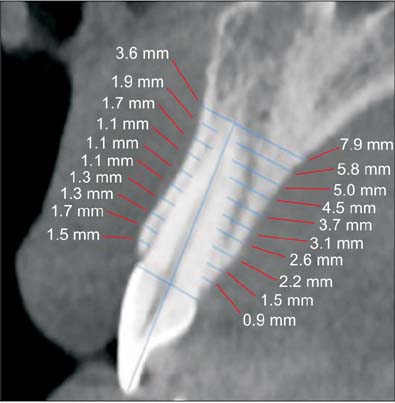
Figure 2 Determination of measurement levels.
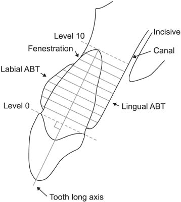
Figure 3 Schematic illustration of labial and lingual bone thickness measurements. Level 0, cementoenamel junction; level 10, root apex area. ABT, Alveolar bone thickness.
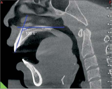
Figure 4 Measuring the angle between the axis of the maxillary central incisors and the palatal plane (U1-PP) in the median sagittal view.
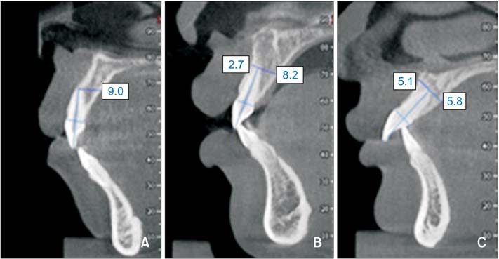
Figure 5 Representative images of three groups classified by different incisor inclination. A, Lingual-inclined group; B, normal group; C, labial-inclined group. The unit of the numberal data in the figure is millimeters.

Figure 6 Fenestration on labial root surface (arrow).
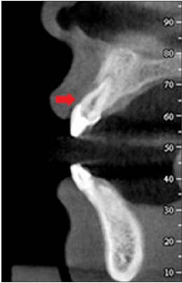
Figure 7 Dehiscence on the root surface (arrow).
Cited by 6 articles
-
Effect of labiolingual inclination of a maxillary central incisor and surrounding alveolar bone loss on periodontal stress: A finite element analysis
Sung-Hwan Choi, Young-Hoon Kim, Kee-Joon Lee, Chung-Ju Hwang
Korean J Orthod. 2016;46(3):155-162. doi: 10.4041/kjod.2016.46.3.155.Quantitative evaluation of midpalatal suture maturation via fractal analysis
Kyoung Ho Kwak, Seong Sik Kim, Yong-Il Kim, Yong-Deok Kim
Korean J Orthod. 2016;46(5):323-330. doi: 10.4041/kjod.2016.46.5.323.Effect of slow forced eruption on the vertical levels of the interproximal bone and papilla and the width of the alveolar ridge
Eun-Young Kwon, Ju-Youn Lee, Jeomil Choi
Korean J Orthod. 2016;46(6):379-385. doi: 10.4041/kjod.2016.46.6.379.Cone-beam computed tomography for the assessment of root–crown ratios of the maxillary and mandibular incisors in a Korean population
Sung-Hwan Choi, Jung-Suk Kim, Cheol-Soon Kim, Hyung-Seog Yu, Chung-Ju Hwang
Korean J Orthod. 2017;47(1):39-49. doi: 10.4041/kjod.2017.47.1.39.Skeletal and dentoalveolar changes after miniscrew-assisted rapid palatal expansion in young adults: A cone-beam computed tomography study
Jung Jin Park, Young-Chel Park, Kee-Joon Lee, Jung-Yul Cha, Ji Hyun Tahk, Yoon Jeong Choi
Korean J Orthod. 2017;47(2):77-86. doi: 10.4041/kjod.2017.47.2.77.Evaluation of changes in the maxillary alveolar bone after incisor intrusion
Ezgi Atik, Hande Gorucu-Coskuner, Bengisu Akarsu-Guven, Tulin Taner
Korean J Orthod. 2018;48(6):367-376. doi: 10.4041/kjod.2018.48.6.367.
Reference
-
1. Handelman CS. The anterior alveolus: its importance in limiting orthodontic treatment and its influence on the occurrence of iatrogenic sequelae. Angle Orthod. 1996; 66:95–109. discussion 109-102. Ten Hoeve A, Mulie RM. The effect of antero-postero incisor repositioning on the palatal cortex as studied with laminagraphy. J Clin Orthod. 1976; 10:804–822.3. Zamora N, Llamas JM, Cibrián R, Gandia JL, Paredes V. Cephalometric measurements from 3D reconstructed images compared with conventional 2D images. Angle Orthod. 2011; 81:856–864.
Article4. Leuzinger M, Dudic A, Giannopoulou C, Kiliaridis S. Root-contact evaluation by panoramic radiography and cone-beam computed tomography of super-high resolution. Am J Orthod Dentofacial Orthop. 2010; 137:389–392.
Article5. de Oliveira AE, Cevidanes LH, Phillips C, Motta A, Burke B, Tyndall D. Observer reliability of three-dimensional cephalometric landmark identification on cone-beam computerized tomography. Oral Surg Oral Med Oral Pathol Oral Radiol Endod. 2009; 107:256–265.
Article6. Ganguly R, Ruprecht A, Vincent S, Hellstein J, Timmons S, Qian F. Accuracy of linear measurement in the Galileos cone beam computed tomography under simulated clinical conditions. Dentomaxillofac Radiol. 2011; 40:299–305.
Article7. Nahm KY, Kang JH, Moon SC, Choi YS, Kook YA, Kim SH, et al. Alveolar bone loss around incisors in Class I bidentoalveolar protrusion patients: a retrospective three-dimensional cone beam CT study. Dentomaxillofac Radiol. 2012; 41:481–488.
Article8. Sarikaya S, Haydar B, Ciğer S, Ariyürek M. Changes in alveolar bone thickness due to retraction of anterior teeth. Am J Orthod Dentofacial Orthop. 2002; 122:15–26.
Article9. Persson RE, Hollender LG, Laurell L, Persson GR. Horizontal alveolar bone loss and vertical bone defects in an adult patient population. J Periodontol. 1998; 69:348–356.
Article10. Lin JX, Xu TM. Contemporary orthodontics. Peking, China: Peking University Medical Press;2011. p. 607.11. Zhou Z, Chen W, Shen M, Sun C, Li J, Chen N. Cone beam computed tomographic analyses of alveolar bone anatomy at the maxillary anterior region in Chinese adults. J Biomed Res. 2014; 28:498–505.
Article12. Andrews LF. Comments on straight-wire appliances. Am J Orthod Dentofacial Orthop. 1990; 98:25A–27A.
Article13. Cao L, Zhang K, Bai D, Jing Y, Tian Y, Guo Y. Effect of maxillary incisor labiolingual inclination and anteroposterior position on smiling profile esthetics. Angle Orthod. 2011; 81:121–129.
Article14. Remmelink HJ, van der. Effects of anteroposterior incisor repositioning on the root and cortical plate: a follow-up study. J Clin Orthod. 1984; 18:42–49.15. Wehrbein H, Bauer W, Diedrich P. Mandibular incisors, alveolar bone, and symphysis after orthodontic treatment. A retrospective study. Am J Orthod Dentofacial Orthop. 1996; 110:239–246.
Article16. Vardimon AD, Graber TM, Voss LR, Lenke J. Determinants controlling iatrogenic external root resorption and repair during and after palatal expansion. Angle Orthod. 1991; 61:113–122. discussion 123-417. Larato DC. Alveolar plate fenestrations and dehiscences of the human skull. Oral Surg Oral Med Oral Pathol. 1970; 29:816–819.
Article18. Larato DC. Alveolar plate defects in children's skulls. J Periodontol. 1972; 43:502.
Article19. Wainwright WM. Faciolingual tooth movement: its influence on the root and cortical plate. Am J Orthod. 1973; 64:278–302.
Article20. Wingard CE, Bowers GM. The effects of facial bone from facial tipping of incisors in monkeys. J Periodontol. 1976; 47:450–454.21. Karring T, Nyman S, Thilander B, Magnusson I. Bone regeneration in orthodontically produced alveolar bone dehiscences. J Periodontal Res. 1982; 17:309–315.
Article22. Patcas R, Müller L, Ullrich O, Peltomäki T. Accuracy of cone-beam computed tomography at different resolutions assessed on the bony covering of the mandibular anterior teeth. Am J Orthod Dentofacial Orthop. 2012; 141:41–50.
Article23. Leung CC, Palomo L, Griffith R, Hans MG. Accuracy and reliability of cone-beam computed tomography for measuring alveolar bone height and detecting bony dehiscences and fenestrations. Am J Orthod Dentofacial Orthop. 2010; 137:4 Suppl. S109–S119.
Article24. Vardimon AD, Oren E, Ben-Bassat Y. Cortical bone remodeling/tooth movement ratio during maxillary incisor retraction with tip versus torque movements. Am J Orthod Dentofacial Orthop. 1998; 114:520–529.
Article25. Barrer HG, Buchin ID, Fogel MS, Swain BF, Ackerman JL. Borderline extraction cases: Panel discussion, part 5. J Clin Orthod. 1971; 5:609–626.26. Ogaard B. Marginal bone support and tooth lengths in 19-year-olds following orthodontic treatment. Eur J Orthod. 1988; 10:180–186.
Article27. Zachrisson BU, Alnaes L. Periodontal condition in orthodontically treated and untreated individuals. II. Alveolar bone loss: radiographic findings. Angle Orthod. 1974; 44:48–55.28. Sun Z, Smith T, Kortam S, Kim DG, Tee BC, Fields H. Effect of bone thickness on alveolar bone-height measurements from cone-beam computed tomography images. Am J Orthod Dentofacial Orthop. 2011; 139:e117–e127.
Article29. Lupi JE, Handelman CS, Sadowsky C. Prevalence and severity of apical root resorption and alveolar bone loss in orthodontically treated adults. Am J Orthod Dentofacial Orthop. 1996; 109:28–37.
Article30. Fuhrmann R. Three-dimensional interpretation of labiolingual bone width of the lower incisors. Part II. J Orofac Orthop. 1996; 57:168–185.31. Wehrbein H, Fuhrmann RA, Diedrich PR. Human histologic tissue response after long-term orthodontic tooth movement. Am J Orthod Dentofacial Orthop. 1995; 107:360–371.
Article
- Full Text Links
-
- Actions
-
Cited
- CITED
-
- Close
- Share
-
- Similar articles
-
- Analysis of the root position of the maxillary incisors in the alveolar bone using cone-beam computed tomography
- Evaluation of changes in the maxillary alveolar bone after incisor intrusion
- Associations among the anterior maxillary dental arch form, alveolar bone thickness, and the sagittal root position of the maxillary central incisors in relation to immediate implant placement: A cone-beam computed tomography analysis
- Analysis of the root position and angulation of maxillary premolars in alveolar bone using cone-beam computed tomography
- Three-dimensional evaluation of maxillary anterior alveolar bone for optimal placement of miniscrew implants
