Cranioplasty Using a Modified Split Calvarial Graft Technique in Cleidocranial Dysplasia
- Affiliations
-
- 1Department of Neurosurgery, Cheju Halla Hospital, Jeju, Korea. hixos@naver.com
- KMID: 2067110
- DOI: http://doi.org/10.3340/jkns.2015.58.1.79
Abstract
- Cleidocranial dysplasia is a well-documented rare autosomal dominant skeletal dysplasia characterized by hypoplastic/aplastic clavicles, brachycephalic skull, patent sutures and fontanelles, midface hypoplasia, and abnormalities of dentition. Patients with cleidocranial dysplasia often complain about undesirable esthetic appearance of their forehead and skull. Notwithstanding many studies of molecular, genetics and skeletal abnormalities of this congenial disorder, there have been very few written reports of cranioplasty involving cleidocranial dysplasia. Thus, we report a rare case of successful cranioplasty using a modified split calvarial graft technique in patient with cleidocranial dysplasia.
Figure
-
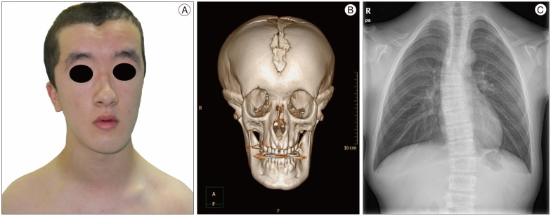
Fig. 1 Note a large head with a midline forehead groove, including a depression of the anterior fontanelle and a depressed nasal bridge, some degree of hypertelorism and a small maxilla (A). A three-dimensional computed tomographic image demonstrating an opened anterior fontanelle, sagittal and metopic suture. Note the high and narrow orbital openings, hypoplasia of the nasal bone and the anteriorly-inclined mandible (B). A chest radiography showing a cone-shaped thorax and hypoplasia of the bilateral clavicles (C).

Fig. 2 The midline calvarial defect areas on the metopic suture and the anterior fontanelle are exposed (A). The bilateral frontal bossing areas are split into the outer and inner table flaps with a thin cutting tool (Midas Rex Legend EHS Stylus High-Speed Surgical Drill, Medtronics). This thin cutting tool can minimize bone losses while splitting the craniotomy flap. The outer table flaps are gained for craniotomy of the calvarial defect area as autografts (B). The bone flour is collected with a bone collector (a widely used generalized sputum collector which was connected to the intraoperative closed suction system) during bone sawing (C and D).
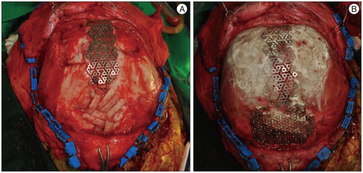
Fig. 3 Note the multiple rectangular-shaped outer tables are put on to the skull defect areas. The areas filled-up with bone flaps and bone flour are covered with a tailored titanium mesh and low profile screws (A). The obtained bone flour and hydroxyapatite cement are molded together in the calvarial defect site. The margins of the bone harvested area are smoothly drilled out with a high speed drill and filled up with hydroxyapatite cement and bone flours for cosmesis (B).

Fig. 4 A post operative frontal photograph of patient. Note the even and flat forehead compared with the preoperative one (A). On the 6 month follow up brain computed tomographic scan demonstrates the bone fusion is noticeable (B).
Reference
-
1. Agrawal A, Garg LN. Split calvarial bone graft for the reconstruction of skull defects. J Surg Tech Case Rep. 2011; 3:13–16. PMID: 22022648.
Article2. Artico M, Ferrante L, Pastore FS, Ramundo EO, Cantarelli D, Scopelliti D, et al. Bone autografting of the calvaria and craniofacial skeleton : historical background, surgical results in a series of 15 patients, and review of the literature. Surg Neurol. 2003; 60:71–79. PMID: 12865021.3. Baumert U, Golan I, Redlich M, Aknin JJ, Muessig D. Cleidocranial dysplasia : molecular genetic analysis and phenotypic-based description of a Middle European patient group. Am J Med Genet A. 2005; 139A:78–85. PMID: 16222673.4. Cooper SC, Flaitz CM, Johnston DA, Lee B, Hecht JT. A natural history of cleidocranial dysplasia. Am J Med Genet. 2001; 104:1–6. PMID: 11746020.
Article5. Inoue A, Satoh S, Sekiguchi K, Ibuchi Y, Katoh S, Ota K, et al. Cranioplasty with split-thickness calvarial bone. Neurol Med Chir (Tokyo). 1995; 35:804–807. PMID: 8657331.6. Kang N, Kim SZ, Jung SN. Correction of depressed forehead with BoneSource in cleidocranial dysplasia. J Craniofac Surg. 2009; 20:564–566. PMID: 19305258.
Article7. Kobayashi S, Uchida K, Baba H, Takeno K, Yayama T, Nakajima H, et al. Atlantoaxial subluxation-induced myelopathy in cleidocranial dysplasia. Case report. J Neurosurg Spine. 2007; 7:243–247. PMID: 17688067.8. McGuire TP, Gomes PP, Lam DK, Sándor GK. Cranioplasty for midline metopic suture defects in adults with cleidocranial dysplasia. Oral Surg Oral Med Oral Pathol Oral Radiol Endod. 2007; 103:175–179. PMID: 17234531.
Article9. Mundlos S. Cleidocranial dysplasia : clinical and molecular genetics. J Med Genet. 1999; 36:177–182. PMID: 10204840.10. Mundlos S, Otto F, Mundlos C, Mulliken JB, Aylsworth AS, Albright S, et al. Mutations involving the transcription factor CBFA1 cause cleidocranial dysplasia. Cell. 1997; 89:773–779. PMID: 9182765.
Article11. O'Broin ES, Morrin M, Breathnach E, Allcutt D, Earley MJ. Titanium mesh and bone dust calvarial patch during cranioplasty. Cleft Palate Craniofac J. 1997; 34:354–356. PMID: 9257028.12. Ronderos JF, Wiles DA, Ragan FA, Dempesy CW, Culicchia FC, Fontana CJ, et al. Cranioplasty using gentamicin-loaded acrylic cement : a test of neurotoxicity. Surg Neurol. 1992; 37:356–360. PMID: 1631760.
Article13. Sahoo NK, Rangan M. Role of split calvarial graft in reconstruction of craniofacial defects. J Craniofac Surg. 2012; 23:e326–e331. PMID: 22801169.
Article14. Strong EB, Moulthrop T. Calvarial bone graft harvest : a new technique. Otolaryngol Head Neck Surg. 2000; 123:547–552. PMID: 11077338.
- Full Text Links
-
- Actions
-
Cited
- CITED
-
- Close
- Share
-
- Similar articles
-
- Cleidocranial Dysostosis: One Case Report
- Sphenoid Dysplasia in the Absence of Neurofibromatosis Type I: Case Report
- Cleidocranial dysplasia: a preliminary report
- Treatment of Fibrous Dysplasia of the Fronto-Orbital Area with Radical Resection and Autogenous Reconstruction Using Split Calvarial Bone Graft: A Case Report
- Cleidocranial Dysostosis One Case Report
