Thoracic Extradural Cavernous Hemangioma Mimicking a Dumbbell-Shaped Tumor
- Affiliations
-
- 1Department of Neurosurgery, Asan Medical Center, University of Ulsan College of Medicine, Seoul, Korea. swroh@amc.seoul.kr
- KMID: 2067108
- DOI: http://doi.org/10.3340/jkns.2015.58.1.72
Abstract
- Dumbbell-shaped spinal extradural cavernous hemangioma is rare. The differential diagnosis of dumbbell-shaped spinal tumors based on magnetic resonance imaging includes schwannoma and lymphoma. Here, we report a dumbbell-shaped spinal extradural cavernous hemangioma with intrathoracic growth on T2-3 in a 64-year-old man complaining of right side infrascapular area back pain with no neurologic deficit. The cavernous hemangioma was resected through combined video-assisted thoracoscopy and laminectomy without a fusion procedure. The patient had tolerable operative wound pain with no neurologic deficit after surgery. Based on magnetic resonance imaging findings and a review of the literature, we discuss cavernous hemangioma among the differential diagnosis of paravertebral dumbbell-shaped spinal tumors and the importance of complete resection.
Keyword
MeSH Terms
Figure
-
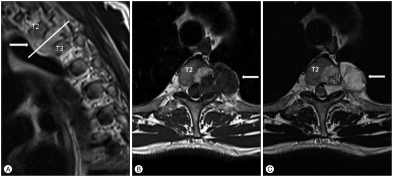
Fig. 1 Preoperative T2-weighted sequence (A and B) and T1-weighted sequence (C) magnetic resonance imaging showing an epidural mass compressing the spinal cord along the T2-3 disc level. The epidural mass revealed iso-signal intensity on T1-weight sequence and high signal intensity on T2-weighted sequence. Preoperative magnetic resonance imaging showed well-demarcated round-shaped abnormal high signal intensity with mottled low signal intensity on both T1- and T2-weighted sequences within the left half of the T2 vertebral body extending to the left pedicle.
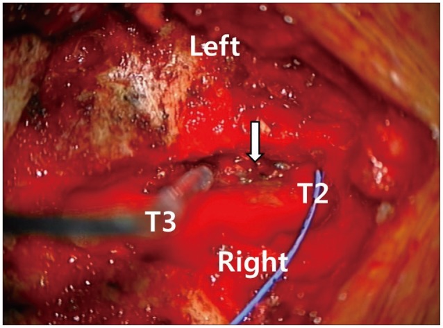
Fig. 2 Intraoperative microscopic photography. Laminectomy was performed from T2 to T4. A pinkish, non-encapsulated mass (white arrow) was found, which was attached to the dura mater and extended through the left T2-3 intervertebral foramen.
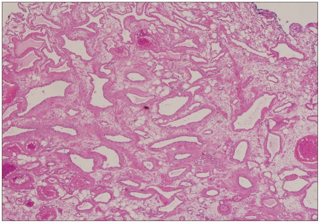
Fig. 3 High-magnification (×40) of a haematoxylin and eosin (H&E) stained section showing the cavernous hemangioma composed of an irregular cavernous vascular space. Some of the vascular spaces contained blood. The walls of the vascular space were lined by a single layer of endothelium.
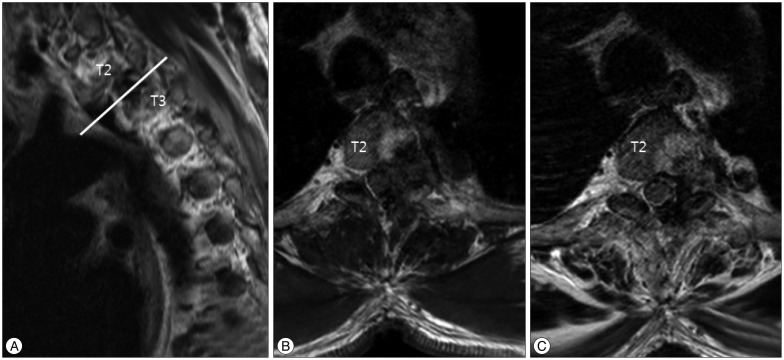
Fig. 4 Postoperative day 1 magnetic resonance images of the patient. T2-weighted sequence (A and B) and T1-weighted sequence (C) images showing no residual extradural cavernous hemangioma. However, the T2 vertebral body cavernous hemagioma was still present.
Cited by 1 articles
-
One Stage Posterior Minimal Laminectomy and Video-Assisted Thoracoscopic Surgery (VATS) for Removal of Thoracic Dumbbell Tumor
Kyoung Hyup Nam, Hyo Yeoung Ahn, Jeong Su Cho, Yeoung Dae Kim, Byung Kwan Choi, In Ho Han
J Korean Neurosurg Soc. 2017;60(2):257-261. doi: 10.3340/jkns.2016.0909.004.
Reference
-
1. A L H, T R, Chamarthy NP, Puri K. A pure epidural spinal cavernous hemangioma - with an innocuous face but a perilous behaviour!! J Clin Diagn Res. 2013; 7:1434–1435. PMID: 23998084.
Article2. Barzin M, Maleki I. Incidence of vertebral hemangioma on spinal magnetic resonance imaging in Northern Iran. Pak J Biol Sci. 2009; 12:542–544. PMID: 19580008.
Article3. Carlier R, Engerand S, Lamer S, Vallee C, Bussel B, Polivka M. Foraminal epidural extra osseous cavernous hemangioma of the cervical spine : a case report. Spine (Phila Pa 1976). 2000; 25:629–631. PMID: 10749642.
Article4. Daneyemez M, Sirin S, Duz B. Spinal epidural cavernous angioma : case report. Minim Invasive Neurosurg. 2000; 43:159–162. PMID: 11108117.5. Demachi H, Takashima T, Kadoya M, Suzuki M, Konishi H, Tomita K, et al. MR imaging of spinal neurinomas with pathological correlation. J Comput Assist Tomogr. 1990; 14:250–254. PMID: 2312854.
Article6. Feider HK, Yuille DL. An epidural cavernous hemangioma of the spine. AJNR Am J Neuroradiol. 1991; 12:243–244. PMID: 1902020.7. Golwyn DH, Cardenas CA, Murtagh FR, Balis GA, Klein JB. MRI of a cervical extradural cavernous hemangioma. Neuroradiology. 1992; 34:68–69. PMID: 1553041.
Article8. Graziani N, Bouillot P, Figarella-Branger D, Dufour H, Peragut JC, Grisoli F. Cavernous angiomas and arteriovenous malformations of the spinal epidural space : report of 11 cases. Neurosurgery. 1994; 35:856–863. discussion 863-864PMID: 7838334.9. Guthkelch AN. Haemangiomas involving the spinal epidural space. J Neurol Neurosurg Psychiatry. 1948; 11:199–210. PMID: 18878025.
Article10. Hong SP, Cho DS, Kim MH, Shin KM. Spinal epidural cavernous hemangioma simulating a disc protrusion: a case report. J Korean Neurosurg Soc. 2003; 33:509–511.11. Lee JP, Wang AD, Wai YY, Ho YS. Spinal extradural cavernous hemangioma. Surg Neurol. 1990; 34:345–351. PMID: 2218857.
Article12. Li TY, Xu YL, Yang J, Wang J, Wang GH. Primary spinal epidural cavernous hemangioma : clinical features and surgical outcome in 14 cases. J Neurosurg Spine. 2015; 22:39–46. PMID: 25343406.
Article13. Luca D, Marta R, Salima M, Valentina B, Domenico D. Spinal epidural cavernous angiomas : a clinical series of four cases. Acta Neurochir (Wien). 2014; 156:283–284. PMID: 24363146.14. Mascalchi M, Torselli P, Falaschi F, Dal Pozzo G. MRI of spinal epidural lymphoma. Neuroradiology. 1995; 37:303–307. PMID: 7666966.
Article15. McCormick WF. The pathology of vascular ("arteriovenous") malformations. J Neurosurg. 1966; 24:807–816. PMID: 5934138.
Article16. Padolecchia R, Acerbi G, Puglioli M, Collavoli PL, Ravelli V, Caciagli P. Epidural spinal cavernous hemangioma. Spine (Phila Pa 1976). 1998; 23:1136–1140. PMID: 9615365.
Article17. Padovani R, Tognetti F, Proietti D, Pozzati E, Servadei F. Extrathecal cavernous hemangioma. Surg Neurol. 1982; 18:463–465. PMID: 7163968.
Article18. Rahman A, Hoque SU, Bhandari PB, Abu Obaida AS. Spinal extradural cavernous haemangioma in an elderly man. BMJ Case Rep. 2012; 2012.
Article19. Saracen A, Kotwica Z. Thoracic spinal epidural cavernous haemangioma with an acute onset : case report and the review of the literature. Clin Neurol Neurosurg. 2013; 115:799–801. PMID: 22918065.
Article20. Saringer W, Nöbauer I, Haberler C, Ungersböck K. Extraforaminal, thoracic, epidural cavernous haemangioma : case report with analysis of magnetic resonance imaging characteristics and review of the literature. Acta Neurochir. 2001; 143(Wien):1293–1297. PMID: 11810396.
Article21. Saumench R, Barcia JA, Arnau A, Cantó A. Combined thoracotomy and laminectomy for spinal cavernomas with intrathoracic growth. Interact Cardiovasc Thorac Surg. 2004; 3:76–78. PMID: 17670181.
Article22. Sharma MS, Borkar SA, Kumar A, Sharma MC, Sharma BS, Mahapatra AK. Thoracic extraosseous, epidural, cavernous hemangioma : case report and review of literature. J Neurosci Rural Pract. 2013; 4:309–312. PMID: 24250167.23. Shin JH, Lee HK, Rhim SC, Park SH, Choi CG, Suh DC. Spinal epidural cavernous hemangioma : MR findings. J Comput Assist Tomogr. 2001; 25:257–261. PMID: 11242225.24. Shivaprasad S, Shroff G, Campbell GA. Thoracic epidural cavernous hemangioma imaging and pathology. JAMA Neurol. 2013; 70:1196–1197. PMID: 23897028.25. Talacchi A, Spinnato S, Alessandrini F, Iuzzolino P, Bricolo A. Radiologic and surgical aspects of pure spinal epidural cavernous angiomas. Report on 5 cases and review of the literature. Surg Neurol. 1999; 52:198–203. PMID: 10447290.
Article26. Yettou H, Vinikoff L, Baylac F, Marchal JC. [Spinal epidural cavernous angioma. Apropos of 2 cases. Review of the literature]. Neurochirurgie. 1996; 42:300–304. discussion 304-305PMID: 9161537.27. Zevgaridis D, Büttner A, Weis S, Hamburger C, Reulen HJ. Spinal epidural cavernous hemangiomas. Report of three cases and review of the literature. J Neurosurg. 1998; 88:903–908. PMID: 9576262.
- Full Text Links
-
- Actions
-
Cited
- CITED
-
- Close
- Share
-
- Similar articles
-
- Two Cases of Epidural Cavernous Hemangioma in the Thoraic Spine
- A Case of Extradural Cavernous Hemangioma with Reuptured Disc
- Epidural Cavernous Hemangioma with Foraminal Extension
- Dumbbell-shaped Epidural Cavernous Hemangioma: A Case Report
- A Dumbbell-shaped Thoraco-lumbar Extradural Ganglioneuroma: Case Report
