Hydrocephalus due to Membranous Obstruction of Magendie's Foramen
- Affiliations
-
- 1Department of Neurosurgery, Athens General Hospital "G. Gennimatas", Athens, Greece. kostaskasapas@gmail.com
- KMID: 2067096
- DOI: http://doi.org/10.3340/jkns.2015.57.1.68
Abstract
- We report a case of non communicating hydrocephalus due to membranous obstruction of Magendie's foramen. A 37-year-old woman presented with intracranial hypertension symptoms caused by the occlusion of Magendie's foramen by a membrane probably due to arachnoiditis. As far as the patient's past medical history is concerned, an Epstein-Barr virus infectious mononucleosis was described. Fundoscopic examination revealed bilateral papilledema. Brain magnetic resonance imaging demonstrated a significant ventricular dilatation of all ventricles and turbulent flow of cerebelospinal fluid (CSF) in the fourth ventricle as well as back flow of CSF through the Monro's foramen to the lateral ventricles. The patient underwent a suboccipital craniotomy with C1 laminectomy. An occlusion of Magendie's foramen by a thickened membrane was recognized and it was incised and removed. We confirm the existence of hydrocephalus caused by fourth ventricle outflow obstruction by a membrane. The nature of this rare entity is difficult to demonstrate because of the complex morphology of the fourth ventricle. Treatment with surgical exploration and incision of the thickened membrane proved to be a reliable method of treatment without the necessity of endoscopic third ventriculostomy or catheter placement.
Keyword
MeSH Terms
Figure
-
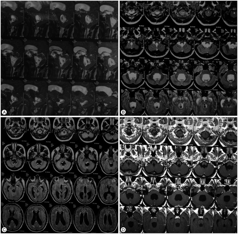
Fig. 1 A : Preoperative sagittal T2W TSE MRI showing turbulent flow of CSF in the fourth ventricle and back flow of CSF through the Monro's foramen to the lateral ventricles. B : Preoperative axial T2 MRI showing significant dilatation of fourth ventricle with turbulent flow of CSF. C : Preoperative axial T1 MRI showing dilatation of third, fourth and lateral ventricles. D : Preoperative axial T1 MRI showing significant dilatation of fourth ventricle. CSF : cerebelospinal fluid.

Fig. 2 A : Perioperative photo showing the occlusion of Magendie's foramen by the membrane. B : Perioperative photo showing Magendie's foramen after the incision and removal of the membrane.
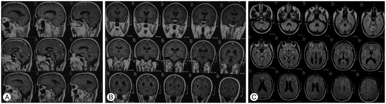
Fig. 3 A : Postoperative sagittal T1 MRI showing marked decrease in the size of the ventricles. B : Postoperative coronal T1 MRI showing marked decrease in the size of the ventricles. C : Postoperative axial T1 MRI showing marked decrease in the size of the ventricles.
Reference
-
1. Amacher AL, Page LK. Hydrocephalus due to membranous obstruction of the fourth ventricle. J Neurosurg. 1971; 35:672–676. PMID: 5315737.
Article2. Hashish H, Guenot M, Mertens P, Sindou M. [Chronic hydrocephalus in an adult due to congenital membranous occlusion of the apertura mediana ventriculi quartii (foramen of Magendie). Report of two cases and review of the literature]. Neurochirurgie. 1999; 45:232–236. PMID: 10567964.3. Holland HC, Graham WL. Congenital atresia of the foramina of Luschka and Magendie with hydrocephalus; report of a case in an adult. J Neurosurg. 1958; 15:688–694. PMID: 13599058.
Article4. Huang YC, Chang CN, Chuang HL, Scott RM. Membranous obstruction of the fourth ventricle outlet. A case report. Pediatr Neurosurg. 2001; 35:43–47. PMID: 11490191.
Article5. Karachi C, Le Guérinel C, Brugières P, Melon E, Decq P. Hydrocephalus due to idiopathic stenosis of the foramina of Magendie and Luschka. Report of three cases. J Neurosurg. 2003; 98:897–902. PMID: 12691419.
Article6. Kim JH, Chung YG, Lee NJ, Kim SH, Lee HK, Lee KC, et al. Unilateral hydrocephalus in congenital atresia of the foramen of Monro. J Korean Neurosurg Soc. 2000; 29:434–437.7. Maloney AF. Two cases of congenital atresia of the foramina of Magendie and Luschka. J Neurol Neurosurg Psychiatry. 1954; 17:134–138. PMID: 13163705.
Article8. Osaka Y, Shin H, Sugawa N, Yoshino E, Horikawa Y, Yamaki T, et al. ["Dis-proportionately large, communicating fourth ventricle" due to membranous obstruction of Magendie's foramen]. No Shinkei Geka. 1995; 23:429–433. PMID: 7753323.9. Rifkinson-Mann S, Sachdev VP, Huang YP. Congenital fourth ventricular midline outlet obstruction. Report of two cases. J Neurosurg. 1987; 67:595–599. PMID: 3309204.
Article10. Rougier A, Ménégon P. MRI evidence of membranous occlusion of the foramen of Magendie. Acta Neurochir. 2009; 151(Wien):693–694. PMID: 19262983.
Article11. van der Klooster JM, van Saase JL, Grootendorst AF, Sinnige HA. [Meningo-encephalitis and hydrocephalus caused by Epstein-Barr virus]. Ned Tijdschr Geneeskd. 1998; 142:650–654. PMID: 9623131.12. Yoshioka S, Matsukado Y, Uemura S, Nagahiro S, Ootsuka T, Yadomi C. [Hydrocephalus due to membranous obstruction of the fourth ventricle aperture]. No Shinkei Geka. 1985; 13:1135–1139. PMID: 4080086.
