Transdural Nerve Rootlet Entrapment in the Intervertebral Disc Space through Minimal Dural Tear : Report of 4 Cases
- Affiliations
-
- 1Department of Neurosurgery, Wooridul Spine Hospital, Seoul, Korea.
- 2Spine Center, Department of Neurosurgery, Seoul St. Mary's Hospital, The Catholic University of Korea College of Medicine, Seoul, Korea. mddavidk@dreamwiz.com
- KMID: 2066987
- DOI: http://doi.org/10.3340/jkns.2013.53.1.52
Abstract
- Four patients underwent lumbar surgery. In all four patients, the dura was minimally torn during the operation. However, none exhibited signs of postoperative cerebrospinal fluid leakage. In each case, a few days after the operation, the patient suddenly experienced severe recurring pain in the leg. Repeat magnetic resonance imaging showed transdural nerve rootlets entrapped in the intervertebral disc space. On exploration, ventral dural tears and transdural nerve rootlet entrapment were confirmed. Midline durotomy, herniated rootlet repositioning, and ventral dural tear repair were performed, and patients' symptoms improved after rootlet repositioning. Even with minimal dural tearing, nerve rootlets may become entrapped, resulting in severe recurring symptoms. Therefore, the dural tear must be identified and repaired during the first operation.
Keyword
Figure
-
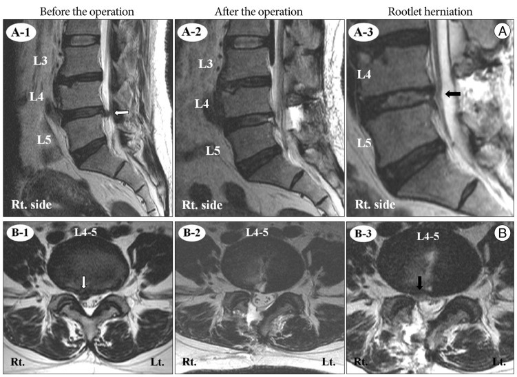
Fig. 1 Before the operation : disc herniation at L4-5 on the right side (white arrow). A-1. Sagittal magnetic resonance image. B-1. Axial magnetic resonance image. After the operation : good decompression of L4-5. A-2. Sagittal magnetic resonance image. B-2. Axial magnetic resonance image. Rootlet herniation : rootlet herniating into the intervertebral disc space at L4-5 on the right side (black arrow). A-3. Sagittal magnetic resonance image. B-3. Axial magnetic resonance image.
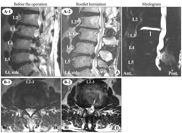
Fig. 2 Before the operation : disc herniation at L2-3, L3-4, and L4-5. A-1. Sagittal magnetic resonance image. B-1. Axial magnetic resonance image showing disc herniation at L2-3 on the left side. Rootlet herniation : rootlet herniating into the intervertebral disc space at L2-3 on the left side (white arrow). A-2. Sagittal magnetic resonance image. B-2. Axial magnetic resonance image of L2-3. Myelogram : magnetic resonance myelogram showing intradiscal leakage of cerebrospinal fluid (white arrow).

Fig. 3 Initial magnetic resonance image : extraforaminal disc herniation at L4-5 on the left side. A-1. Sagittal magnetic resonance image. B-1. Axial magnetic resonance image of L4-5. Before the operation : paramedian disc herniation and extraforaminal disc herniation at L4-5 on the left side. A-2. Sagittal magnetic resonance image. B-2. Axial magnetic resonance image of L4-5. Rootlet herniation : rootlet herniating into the intervertebral disc space at L4-5 on the left side (white arrow). A-3. Sagittal magnetic resonance image. B-3. Axial magnetic resonance image of L4-5.

Fig. 4 Before the operation : disc herniation with osteophyte at L1-2 (white arrow). A-1. Sagittal magnetic resonance image. B-1. Axial magnetic resonance image of L1-2. Rootlet herniation : rootlet herniating into the intervertebral disc space at L1-2 (white arrow). After the operation : herniated rootlets were reposited.
Cited by 1 articles
-
The Incidence and Management of Dural Tears and Cerebrospinal Fluid Leakage during Corrective Osteotomy for Ankylosing Spondylitis with Kyphotic Deformity
Dae-Jean Jo, Ki-Tack Kim, Sang-Hun Lee, Myung-Guk Cho, Eun-Min Seo
J Korean Neurosurg Soc. 2015;58(1):60-64. doi: 10.3340/jkns.2015.58.1.60.
Reference
-
1. Asha MJ, George KJ, Choksey M. Pseudomeningocele presenting with cauda equina syndrome : is a 'ball-valve' theory the answer? Br J Neurosurg. 2011; 25:766–768. PMID: 21591855.
Article2. Aydinli U, Karaeminoğullari O, Tişkaya K, Oztürk C. Dural tears in lumbar burst fractures with greenstick lamina fractures. Spine (Phila Pa 1976). 2001; 26:E410–E415. PMID: 11547211.3. Cammisa FP Jr, Eismont FJ, Green BA. Dural laceration occurring with burst fractures and associated laminar fractures. J Bone Joint Surg. 1989; 71:1044–1052. PMID: 2760080.
Article4. Cammisa FP Jr, Girardi FP, Sangani PK, Parvataneni HK, Cadag S, Sandhu HS. Incidental durotomy in spine surgery. Spine (Phila Pa 1976). 2000; 25:2663–2667. PMID: 11034653.
Article5. Couture D, Branch CL Jr. Spinal pseudomeningoceles and cerebrospinal fluid fistulas. Neurosurg Focus. 2003; 15:E6. PMID: 15305842.
Article6. Eismont FJ, Wiesel SW, Rothman RH. Treatment of dural tears associated with spinal surgery. J Bone Joint Surg Am. 1981; 63:1132–1136. PMID: 7024283.
Article7. Hasegawa K, Yamamoto N. Nerve root herniation secondary to lumbar puncture in the patient with lumbar canal stenosis. A case report. Spine (Phila Pa 1976). 1999; 24:915–917. PMID: 10327516.
Article8. Hodges SD, Humphreys SC, Eck JC, Covington LA. Management of incidental durotomy without mandatory bed rest. A retrospective review of 20 cases. Spine (Phila Pa 1976). 1999; 24:2062–2064. PMID: 10528385.9. Hosono N, Yonenobu K, Ono K. Postoperative cervical pseudomeningocele with herniation of the spinal cord. Spine (Phila Pa 1976). 1995; 20:2147–2150. PMID: 8588173.
Article10. Khan MH, Rihn J, Steele G, Davis R, Donaldson WF 3rd, Kang JD, et al. Postoperative management protocol for incidental dural tears during degenerative lumbar spine surgery : a review of 3,183 consecutive degenerative lumbar cases. Spine (Phila Pa 1976). 2006; 31:2609–2613. PMID: 17047553.
Article11. Lee KS, Hardy IM 2nd. Postlaminectomy lumbar pseudomeningocele : report of four cases. Neurosurgery. 1992; 30:111–114. PMID: 1738437.12. Nishi S, Hashimoto N, Takagi Y, Tsukahara T. Herniation and entrapment of a nerve root secondary to an unrepaired small dural laceration at lumbar hemilaminectomies. Spine (Phila Pa 1976). 1995; 20:2576–2579. PMID: 8610254.
Article13. O'Connor D, Maskery N, Griffiths WE. Pseudomeningocele nerve root entrapment after lumbar discectomy. Spine (Phila Pa 1976). 1998; 23:1501–1502. PMID: 9670405.14. Park DK, Khurana N, Ellman MB, Singh K. Perioperative complications in lumbar spine surgery. Contemp Spine Surg. 2009; 10:1–6.
Article15. Pavlou G, Bucur SD, van Hille PT. Entrapped spinal nerve roots in a pseudomeningocoele as a complication of previous spinal surgery. Acta Neurochir (Wien). 2006; 148:215–219. discussion 219-220. PMID: 16374564.
Article16. Saxler G, Krämer J, Barden B, Kurt A, Pförtner J, Bernsmann K. The long-term clinical sequelae of incidental durotomy in lumbar disc surgery. Spine (Phila Pa 1976). 2005; 2298–2302. PMID: 16227893.
Article17. Töppich HG, Feldmann H, Sandvoss G, Meyer F. Intervertebral space nerve root entrapment after lumbar disc surgery Two cases. Spine (Phila Pa 1976). 1994; 249–250. PMID: 8153836.
- Full Text Links
-
- Actions
-
Cited
- CITED
-
- Close
- Share
-
- Similar articles
-
- Symptomatic Epidural Gas-containing Cyst from Intervertebral Vacuum Phenomenon
- Clinical and Radiological Findings of Nerve Root Herniation after Discectomy of Lumbar Disc Herniation
- Relationship between Lamina Fractures and Dural Tear in Low Lumbar Burst Fractures
- Functional myelography
- Experience with Microsurgery in the Herniations of Lumbar, Cervical and Thoracic Intervertebral Discs
