The implant overdenture on the edentulous mandible using CAD/CAM system: A case report
- Affiliations
-
- 1Department of Prosthodontics, School of Dentistry, Pusan National University, Yangsan, Republic of Korea. huhjb@pusan.ac.kr
- KMID: 2057209
- DOI: http://doi.org/10.4047/jkap.2015.53.1.66
Abstract
- Alveolar bone loss and deformation can be a risk factor in removable prosthetic restoration treatment for partially or fully edentulous patients. The use of implants to solve this problem could improve the support, retention and stability of removable restoration. Attachments used in implant overdenture are versatile. The attachment should be selected according to the patients' conditions. Milled bar has been chosen when readymade bar could not be used because of the narrow distance between implants or firm stability and support of supra-structure were needed. Milled bar design is able to provide cross arch stabilization and comfortability to patients. However, it needs skilled laboratory procedures. Recently, the fabrication of milled bar has become simple and its suitability has been improved through the development of CAD/CAM system. In a 67-year-old female Alzheimer's disease patient with 8 implant fixtures on the fully edentulous site of mandible, implant overdenture with using milled bar and magnet attachment was planned. As rapid treatment was required, CAD/CAM system was used to make a simple laboratory procedure instead of a traditional fabrication process. With this system, implant overdenture with milled bar can be fabricated esthetically and functionally.
MeSH Terms
Figure
-

Fig. 1. Intraoral view of the patient's preoperative condition. (A, E) Occlusal view of maxilla and mandible, (B, D) Lateral view, (C) Frontal view.

Fig. 2. Final impression taking of maxilla and mandible (B, E) with individual trays (A, D) and Definitive cast fabrication of maxilla and mandible (C, F).

Fig. 3. Laboratory procedures using 3Shape software. (A) Scanned customized abutments (Gray), gingiva (Pink) and bite material of antagonistic teeth (Purple), (B) Confirmation of fixture and hole direction, (C) Mandibular teeth arrangement according to the maxillary temporary denture, (D) Establishment of bar margin, (E) Design a milled bar and decision the position of magnet attachments, (F) Evaluation of the bar position according to the arranged and antagonistic teeth.
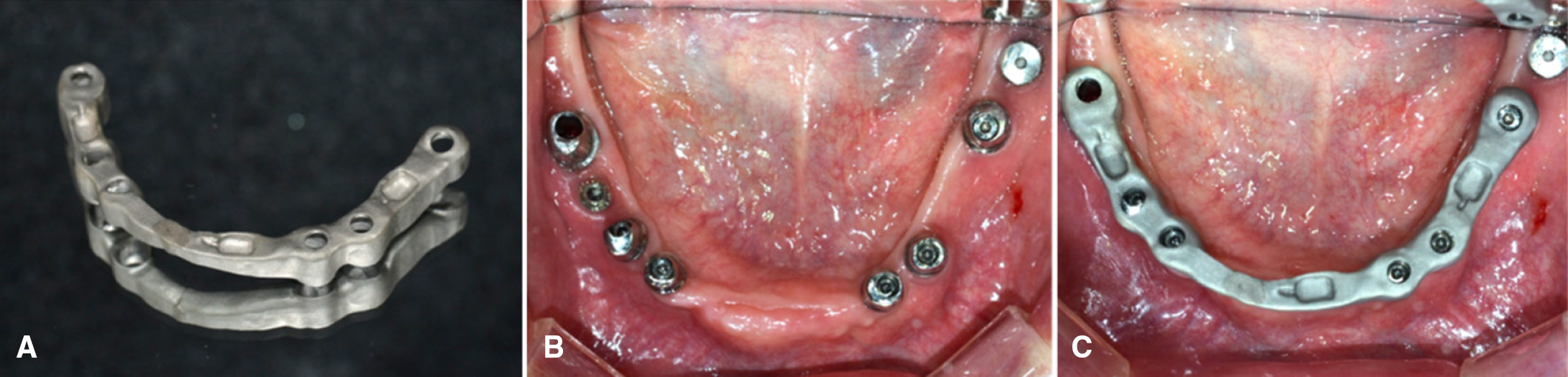
Fig. 4. Fabricated milled bar (A) and verification of bar position in the oral cavity (B, C).

Fig. 5. Decision of vertical dimension using Wilis method (A, B), Facebow transfer (C) and mounted master casts according to the jaw relation record (D).
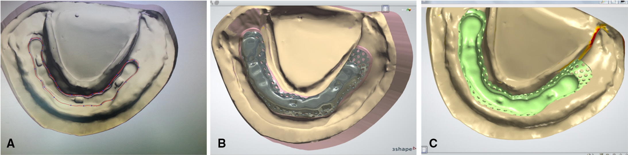
Fig. 6. Framework design. covering milled bar (A), giving the autopolymerizing acrylic resin space under the framework (pink) (B), and confirmation of magnet position on the framework using CAD/CAM system (C).
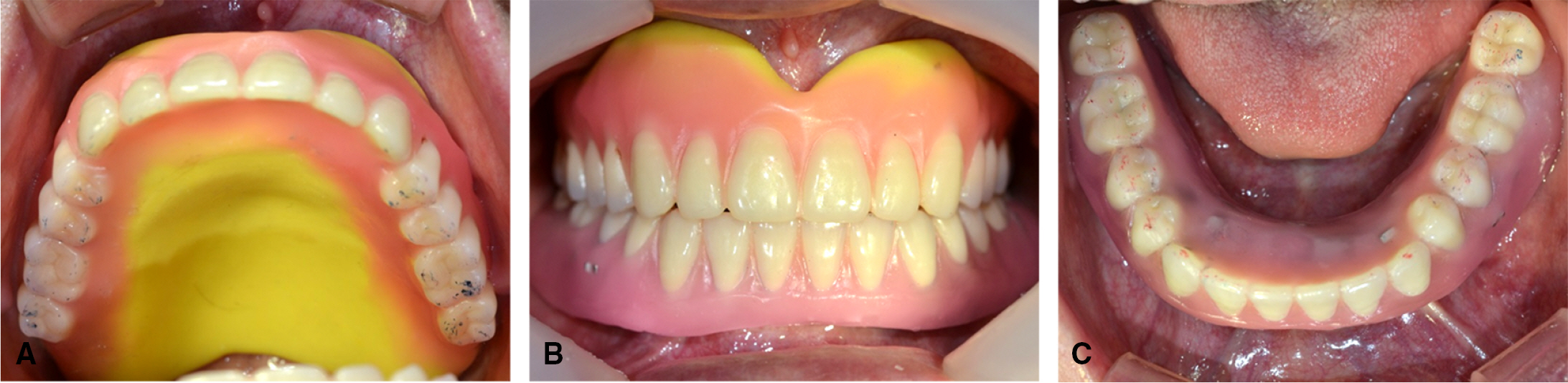
Fig. 7. Esthetic try-in with wax denture. (A, C) Occlusal view of maxilla and mandible, (B) Frontal view.
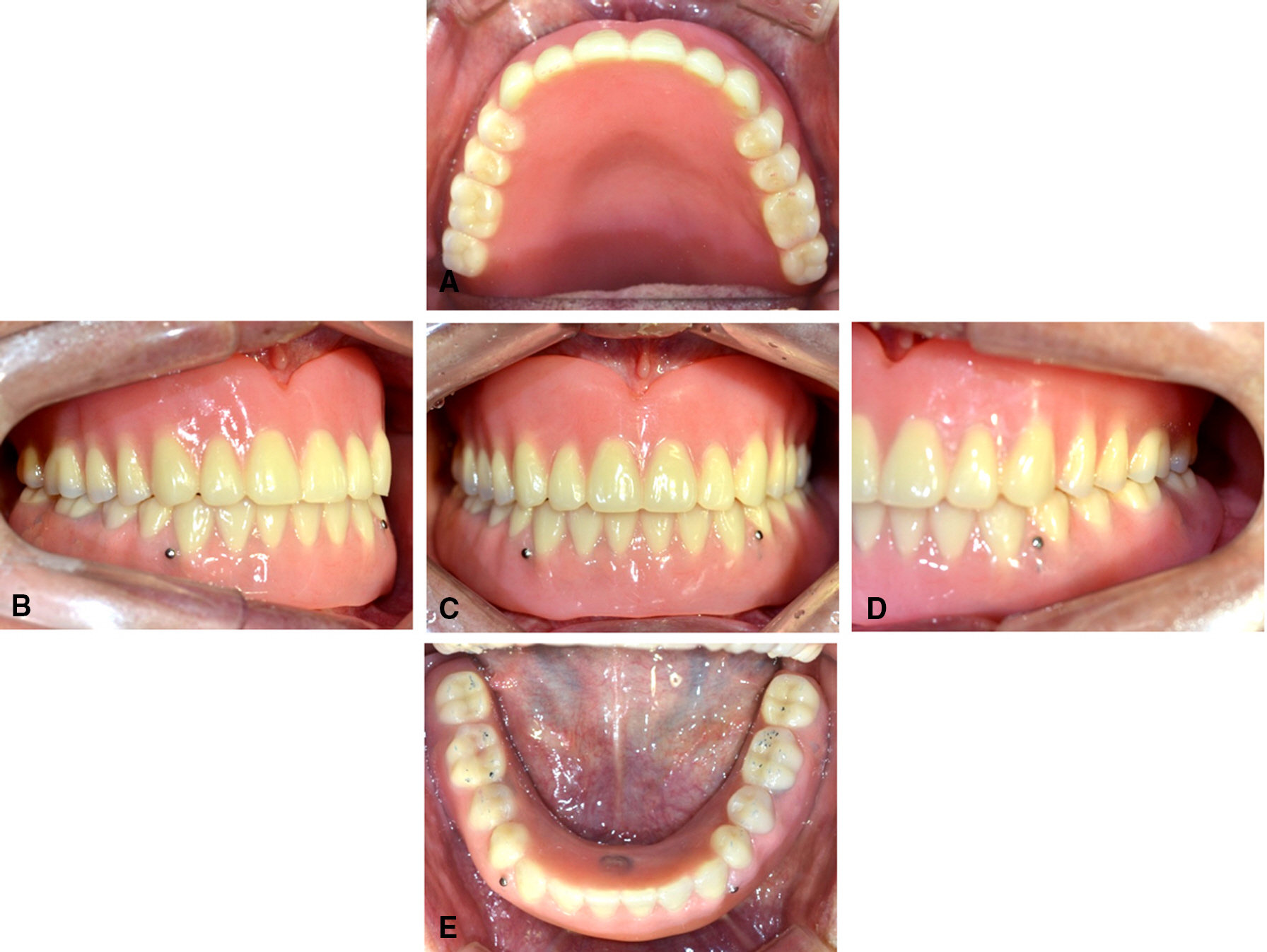
Fig. 8. Final restoration was placed in patient's oral cavity. (A, E) Occlusal view of maxilla and mandible, (B, D) Lateral view, (C) Frontal view.
Reference
-
1. Kim SW. Implant overdenture. Seoul: MyoungMun Publishing Co;2007.2. Bra�nemark PI, Adell R, Breine U, Hansson BO, Lindstro¨m J, Ohlsson A. Intra-osseous anchorage of dental prostheses. I. Experimental studies. Scand J Plast Reconstr Surg. 1969; 3:81–100.3. DeBoer J. Edentulous implants: overdenture versus fixed. J Prosthet Dent. 1993; 69:386–90.
Article4. Trakas T, Michalakis K, Kang K, Hirayama H. Attachment systems for implant retained overdentures: a literature review. Implant Dent. 2006; 15:24–34.
Article5. Sadowsky SJ. The implant-supported prosthesis for the edentulous arch: design considerations. J Prosthet Dent. 1997; 78:28–33.
Article6. Adell R, Lekholm U, Rockler B, Bra�nemark PI. A 15-year study of osseointegrated implants in the treatment of the edentulous jaw. Int J Oral Surg. 1981; 10:387–416.
Article7. Zarb GA, Bolender CL, Eckert SE, Fenton AH, Jacob RF, Mericske-Stern R. Prosthodontic treatment for edentulous patients: Complete dentures and implant-supported prostheses. 12th ed.Mosby;2003.8. Duret F, Preston JD. CAD/CAM imaging in dentistry. Curr Opin Dent. 1991; 1:150–4.9. Miyazaki T, Hotta Y, Kunii J, Kuriyama S, Tamaki Y. A review of dental CAD/CAM: current status and future perspectives from 20 years of experience. Dent Mater J. 2009; 28:44–56.
Article10. Mo¨rmann WH, Brandestini M, Lutz F, Barbakow F. Chairside computer-aided direct ceramic inlays. Quintessence Int. 1989; 20:329–39.11. Beuer F, Schweiger J, Edelhoff D. Digital dentistry: an overview of recent developments for CAD/CAM generated restorations. Br Dent J. 2008; 204:505–11.
Article12. Persson A, Andersson M, Oden A, Sandborgh-Englund G. A three-dimensional evaluation of a laser scanner and a touch-probe scanner. J Prosthet Dent. 2006; 95:194–200.
Article13. Vlaar ST, van der Zel JM. Accuracy of dental digitizers. Int Dent J. 2006; 56:301–9.
Article14. Rudolph H, Luthardt RG, Walter MH. Computer-aided analysis of the influence of digitizing and surfacing on the accuracy in dental CAD/CAM technology. Comput Biol Med. 2007; 37:579–87.
Article15. Kim KB, Kim JH, Kim WC, Kim JH. Three-dimensional evaluation of gaps associated with fixed dental prostheses fabricated with new technologies. J Prosthet Dent. 2014; 112:1432–6.
Article16. Noack F. CoCr-Revolution. Dent Dialog. 2012; 13:2–5.17. Becker JJ. Permanent magnets. Sci American. 1970; 233:92–100.
Article18. Strnat KJ. The hard magnetic properties of rare earth-transition metal alloys. IEEE Trans Magn. 1972; 8:511–6.19. Sagawa M, Fujimura S, Yamamoto H, Matsuura Y, Hiraga K. Permanent magnet materials based on the rare earth-iron-boron tetragonal compounds. IEEE Trans Magn. 1984; 20:1584–9.
Article20. Sagawa M, Fujimura S, Togawa N, Yamamoto H, Matsuura Y. New material for permanent magnets on a base of Nd and Fe. J Appl Phys. 1984; 55:2083–7.21. Riley MA, Walmsley AD, Harris IR. Magnets in prosthetic dentistry. J Prosthet Dent. 2001; 86:137–42.
Article22. Ceruti P, Bryant SR, Lee JH, MacEntee MI. Magnet-retained implant-supported overdentures: review and 1-year clinical report. J Can Dent Assoc. 2010; 76:a52.23. Smith GA, Laird WR, Grant AA. Magnetic retention units for overdentures. J Oral Rehabil. 1983; 10:481–8.
Article
- Full Text Links
-
- Actions
-
Cited
- CITED
-
- Close
- Share
-
- Similar articles
-
- Fabrication of maxillary complete denture and mandibular implant retained overdenture using CAD-CAM system and Monolithic disc: a case report
- Rehabilitation using mandibular implant overdenture with CAD/CAM milled bar: A case report
- Rehabilitation of edentulous maxilla with implant-supported milled bar overdenture using CAD/CAM customized abutment: A case report
- Mandibular implant-supported overdenture using CAD-CAM Konus type attachment: A case report
- Detachable zirconia prosthesis using Milled bar and ADDTOC attachment in partial edentulous mandible: A case report
