Hip Pelvis.
2014 Sep;26(3):202-205. 10.5371/hp.2014.26.3.202.
Primary Aneurysmal Bone Cyst in the Iliac Bone: A Case Report
- Affiliations
-
- 1Department of Orthopaedic Surgery, St. Carollo Hospital, Suncheon, Korea.
- 2Department of Orthopaedic Surgery, Wonkwang University College of Medicine, Iksan, Korea. osksh@wku.ac.kr
- KMID: 2054187
- DOI: http://doi.org/10.5371/hp.2014.26.3.202
Abstract
- Symptomatic aneurysmal bone cysts with expansible lesions in the pelvis are rare in children. The management of an aggressive vascular lesion in a female child is challenging. The standard treatment for aneurysmal bone cysts is accompanied by a high risk of local recurrence. A 12-year-old female presented with a history of pelvic pain for 5 months. Plain radiographs and magnetic resonance imaging showed a very large expansile lytic lesion arising from the right iliac bone. Intralesional curettage, electric cauterization, chemical sclerotherapy and allogeneic bone graft were performed through the window of the iliac crest. At a follow-up consultation 3.5 years post-surgery, the child had painless full-range movement in the hip joint with no recurrence. Although many treatment options are described, our patient was treated successfully using curettage and allogeneic bone graft without recurrence.
Keyword
MeSH Terms
Figure
-

Fig. 1 Pre-operative anteroposterior radiograph of the pelvis showing an expansile osteolytic lesion involving to the superior border of the acetabulum with multiple septation.
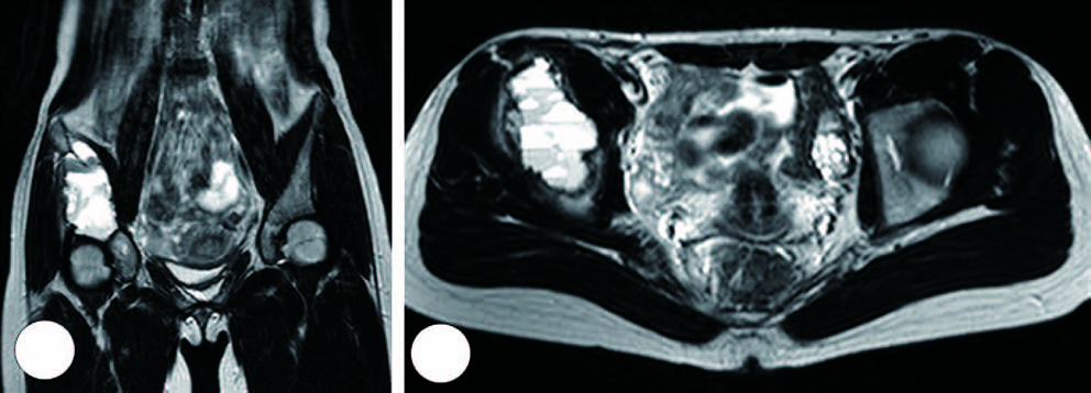
Fig. 2 T2-weighted magnetic resonance imaging coronal view (A) shows 11×9×5 cm large, well defined lesion and axial view (B) shows multiseptations forming cysts containing fluid-like signal intensity.
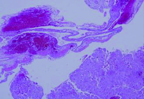
Fig. 3 Histological features composed of hemorrhagic spaces of blood-filled cavities surrounded by fibrotic septa, inflammatory cells and osteoclast cells that are distributed around the cystic spaces (H&E stain, 40×).

Fig. 4 Follow-up radiograph at 3 years and 6 months post-surgery. The radiograph shows good remodeling and no involvement of the hip joint.
Reference
-
1. Papagelopoulos PJ, Choudhury SN, Frassica FJ, Bond JR, Unni KK, Sim FH. Treatment of aneurysmal bone cysts of the pelvis and sacrum. J Bone Joint Surg Am. 2001; 83-A:1674–1681.
Article2. Campanacci M, Capanna R, Picci P. Unicameral and aneurysmal bone cysts. Clin Orthop Relat Res. 1986; 204:25–36.
Article3. Rossi G, Rimondi E, Bartalena T, et al. Selective arterial embolization of 36 aneurysmal bone cysts of the skeleton with N-2-butyl cyanoacrylate. Skeletal Radiol. 2010; 39:161–167.
Article4. Al-Qattan MM. Bipolar electric cauterization as adjuvant treatment after curettage of aneurysmal bone cysts of the hand. Ann Plast Surg. 2014; 72:38–40.
Article5. Cottalorda J, Kohler R, Chotel F, et al. Recurrence of aneurysmal bone cysts in young children: a multicentre study. J Pediatr Orthop B. 2005; 14:212–218.
Article6. Rossi G, Mavrogenis AF, Papagelopoulos PJ, Rimondi E, Ruggieri P. Successful treatment of aggressive aneurysmal bone cyst of the pelvis with serial embolization. Orthopedics. 2012; 35:e963–e968.
Article7. Lambot-Juhan K, Pannier S, Grévent D, et al. Primary aneurysmal bone cysts in children: percutaneous sclerotherapy with absolute alcohol and proposal of a vascular classification. Pediatr Radiol. 2012; 42:599–605.
Article8. Mason KP, Michna E, Zurakowski D, Koka BV, Burrows PE. Serum ethanol levels in children and adults after ethanol embolization or sclerotherapy for vascular anomalies. Radiology. 2000; 217:127–132.
Article9. Agarwal A, Goel P, Khan SA, Kumar P, Qureshi NA. Large aneurysmal bone cyst of iliac bone in a female child: a case report. J Orthop Surg Res. 2010; 5:24.
Article10. Sharifah M, Nurhazla H, Suraya A, Tan S. Pelvic aneurysmal bone cyst. Biomed Imaging Interv J. 2011; 7:e24.
