Obstet Gynecol Sci.
2014 Sep;57(5):415-418. 10.5468/ogs.2014.57.5.415.
Primary omental torsion diagnosed during hysterectomy
- Affiliations
-
- 1Department of Obstetrics and Gynecology, Jeju National University School of Medicine, Jeju, Korea. kimsy@jejunu.ac.kr
- KMID: 2051727
- DOI: http://doi.org/10.5468/ogs.2014.57.5.415
Abstract
- Omental torsion is a rare cause of acute abdominal pain. It presents with nonspecific symptoms and signs of an acute abdomen, making it difficult to diagnose preoperatively, because symptoms mimic those caused by other conditions such as appendicitis, cholecystitis, diverticulitis, and other gynecologic diseases. Computed tomography is an effective and useful method to diagnose and exclude other acute abdominal conditions. Our case presented with sudden right upper abdominal pain with tenderness, rebound tenderness, mild fever (37.2degrees C), increased erythrocyte sedimentation rate (37 mm/hr), increased high-sensitivity C-reactive protein level (5.97 mg/dL). Computed tomography showed a large, well-circumscribed heterogeneous fatty mass and a 7.3 cm subserosal myoma. We could not exclude the myoma as the cause of acute abdominal pain, so we performed an emergency operation with suspicion of omental torsion or necrotic degeneration of the myoma. During the operation, we diagnosed primary omental torsion with infarction and subserosal myoma without secondary degeneration.
Keyword
MeSH Terms
Figure
-

Fig. 1 Axial computerized tomography scan shows a fat pattern characteristic of omental torsion. The vascular pedicle extends caudally and enters a large well-circumscribed heterogeneous fatty mass in the right upper quadrant, and fat density is increased.
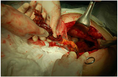
Fig. 2 During the operation, a 10.0×8.0-cm dark brown clockwise omental torsion was observed in the right side of the greater omentum.
Reference
-
1. Bush P. A case of hæmorrhage into the great omentum. Lancet. 1896; 147:286.2. Eitel GG. Rare omental torsion. NY Med Rec. 1899; 55:715–716.3. Basson SE, Jones PA. Primary torsion of the omentum. Ann R Coll Surg Engl. 1981; 63:132–134.4. Adams JT. Torsion of the omentum: abdominal wall, omentum, mesentery and retroperitoneum. In : Schwartz SI, Shires GT, Spencer FC, editors. Principles of surgery. 5th ed. New York: McGraw-Hill;1989. p. 1495–1496.5. Tsironis A, Zikos N, Bali C, Pappas-Gogos G, Koulas S, Katsamakis N. Primary torsion of the greater omentum: report of two cases and review of the literature. Int J Surg. 2007; 17:12884.6. Puylaert JB. Right-sided segmental infarction of the omentum: clinical, US, and CT findings. Radiology. 1992; 185:169–172.7. Epstein LI, Lempke RE. Primary idiopathic segmental infarction of the greater omentum: case report and collective review of the literature. Ann Surg. 1968; 167:437–443.8. Theriot JA, Sayat J, Franco S, Buchino JJ. Childhood obesity: a risk factor for omental torsion. Pediatrics. 2003; 112(6 Pt 1):e460.9. Van Breda Vriesman AC, Lohle PN, Coerkamp EG, Puylaert JB. Infarction of omentum and epiploic appendage: diagnosis, epidemiology and natural history. Eur Radiol. 1999; 9:1886–1892.10. Young TH, Lee HS, Tang HS. Primary torsion of the greater omentum. Int Surg. 2004; 89:72–75.11. Andreuccetti J, Ceribelli C, Manto O, Chiaretti M, Negro P, Tuscano D. Primary omental torsion (POT): a review of literature and case report. World J Emerg Surg. 2011; 6:6.12. Karayiannakis AJ, Polychronidis A, Chatzigianni E, Simopoulos C. Primary torsion of the greater omentum: report of a case. Surg Today. 2002; 32:913–915.13. Kim J, Kim Y, Cho OK, Rhim H, Koh BH, Kim YS, et al. Omental torsion: CT features. Abdom Imaging. 2004; 29:502–504.14. Costi R, Cecchini S, Randone B, Violi V, Roncoroni L, Sarli L. Laparoscopic diagnosis and treatment of primary torsion of the greater omentum. Surg Laparosc Endosc Percutan Tech. 2008; 18:102–105.15. Nubi A, McBride W, Stringel G. Primary omental infarct: conservative vs operative management in the era of ultrasound, computerized tomography, and laparoscopy. J Pediatr Surg. 2009; 44:953–956.
