Primary Intracranial Fibrosarcoma Presenting with Hemorrhage
- Affiliations
-
- 1Department of Neurosurgery, Konkuk University Medical Center, Seoul, Korea. kohyc@kuh.ac.kr
- 2Department of Radiology, Konkuk University Medical Center, Seoul, Korea.
- 3Department of Pathology, Konkuk University Medical Center, Seoul, Korea.
- KMID: 2048481
- DOI: http://doi.org/10.14791/btrt.2013.1.2.91
Abstract
- Primary intracranial fibrosarcomas (PIFs) are extremely rare and the origin of these tumors is still controversial. The rarity of primary intracranial fibrosarcomas makes it difficult to diagnose them correctly and establish a standard treatment. The pathologic diagnosis is made by distinguishing findings from light microscopic and immunohistochemistry analysis. PIFs have been known to be very aggressive neoplasms. The extra-axial location of the tumor could provide an opportunity to perform a total resection even if it does not mean a cure. We present a case of PIFs mimicking a falx meningioma in a 17-year-old man.
MeSH Terms
Figure
-

Fig. 1 Large mass with hemorrhage originating from the falx cerebi (A: Non-enhanced CT, B: T2-weighted MR), ACA was encapsulated by the tumor (white arrow). Well-enhancing tumor with hemorrhage and suspicious brain parenchyma invasion on gadolinium enhancement (C: Axial view, D: Coronal view). Large hypervascular mass in both frontal areas fed by pial branches of both ACAs. Multiple aneurysm, ecstatic changes and luminal irregularities in the A2-3 junction (E: Angiography, F: Angiography 3D). CT: computed tomography, MR: magnetic resonance, ACA: anterior cerebral artery, 3D: three dimension.
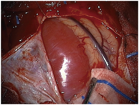
Fig. 2 Well-demarcated hypervascular tumor.
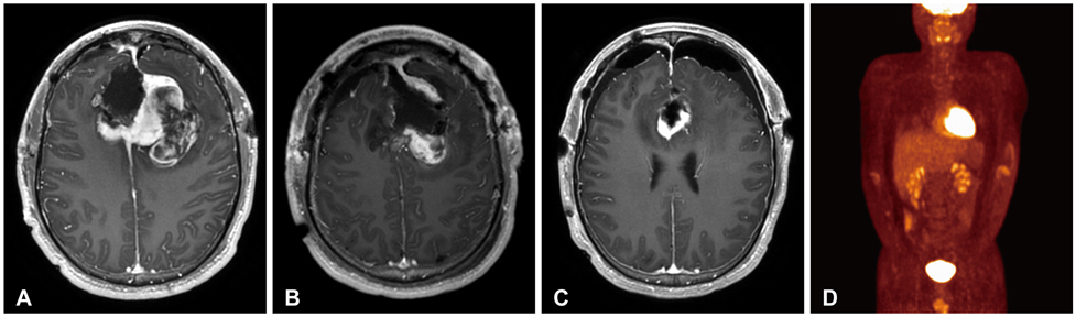
Fig. 3 Images of MRI and PET-CT. A: Residual tumor in the bifrontal region (1st stage post-operation). B: Further resection of residual tumor (2nd stage post-operation). C: Small residual tumor in the anterior pericallosal region (3rd stage post-operation). D: No significant abnormal hypermetabolic lesion suggesting malignancy except brain. PET-CT: positron emission tomography-CT.
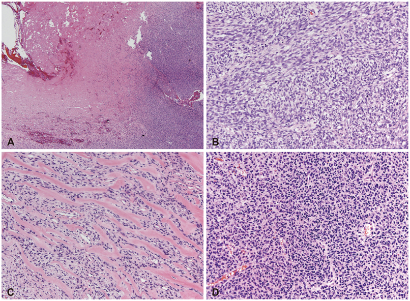
Fig. 4 Histologic features of tumor. A: Tumor based on the dura (H&E, ×40). B: Malignant spindle cell proliferation with herringbone and fascicular pattern (H&E, ×200). C: Tumor with abundant extracellular collagenous matrix production (H&E, ×200). D: Focal presence of malignant small round cell features (H&E, ×200).
Reference
-
1. Chopra R, Bhardwaj M, Premsagar IC. Fibrosarcoma of the meninges. Rare Tumors. 2010; 2:e3.
Article2. Torres G, Petit F, Vilchez V, et al. Primary cerebral fibrosarcoma in a child. Clin Neuropathol. 2007; 26:284–287.
Article3. Bisogno G, Roganovic J, Carli M, et al. Primary intracranial fibrosarcoma. Childs Nerv Syst. 2002; 18:648–651.
Article4. McDonald P, Guha A, Provias J. Primary intracranial fibrosarcoma with intratumoral hemorrhage: neuropathological diagnosis with review of the literature. J Neurooncol. 1997; 35:133–139.5. Vatsal DK, Sharma S, Renjen PN, Kaul S, Jha AN. Primary fibrosarcoma of brain. Neurol India. 2000; 48:396–398.6. Paulus W, Slowik F, Jellinger K. Primary intracranial sarcomas: histopathological features of 19 cases. Histopathology. 1991; 18:395–402.
Article7. Donnet A, Figarella-Branger D, Grisoli F. Primary meningeal fibrosarcoma: a particular neuroradiological presentation. J Neurooncol. 1999; 42:79–83.8. Cai N, Kahn LB. A report of primary brain fibrosarcoma with literature review. J Neurooncol. 2004; 68:161–167.
Article
