Heterogeneous living donor hepatic fat distribution on MRI chemical shift imaging
- Affiliations
-
- 1Department of Surgery, Seoul National University Bundang Hospital, Seongnam, Korea.
- 2Department of Radiology, Seoul National University College of Medicine, Seoul, Korea.
- 3Department of Surgery, Seoul National University College of Medicine, Seoul, Korea. kssuh@snu.ac.kr
- KMID: 2047712
- DOI: http://doi.org/10.4174/astr.2015.89.1.37
Abstract
- PURPOSE
We evaluated the heterogeneity of steatosis in living donor livers to determine its regional differences.
METHODS
Between June 2011 and February 2012, 81 liver donors were selected. Fat fraction was estimated using magnetic resonance triple-echo chemical shifting gradient imaging in 13 different regions: segment 1 (S1), S2, S3, and each peripheral and deep region of S4, S5, S6, S7, and S8.
RESULTS
There were differences (range, 3.2%-5.3%) in fat fractions between each peripheral and deep region of S4, S6, S7, and S8 (P < 0.001, P = 0.004, P < 0.001, and P = 0.006). Fat deposit amount in S1, S2, S3 and deep regions of S4-S8 were significantly different from one another (F [4.003, 58.032] = 8.684, P < 0.001), while there were no differences among the peripheral regions of S4-S8 (F [2.9, 5.3] = 1.3, P = 0.272) by repeated measure analysis of variance method. And regional differences of the amount of fat deposit in the whole liver increased as a peripheral fat fraction of S5 increased (R2 = 0.428, P < 0.001).
CONCLUSION
Multifocal fat measurements for the whole liver are needed because a small regional evaluation might not represent the remaining liver completely, especially in patients with severe hepatic steatosis.
MeSH Terms
Figure
-
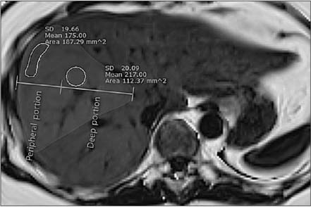
Fig. 1 Estimating the fat fraction in peripheral region and deep region. The fat fraction (1/10 of the mean value in the region of interest) in the peripheral region measures 17.5% and in the deep region measures 21.7%. SD, standard deviation.
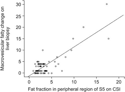
Fig. 2 Linear regression between peripheral fat fraction in segment 5 (S5) on CSI and macrovesicular fatty change on liver biopsy (R2 = 0.667). CSI, magnetic resonance chemical shift imaging.
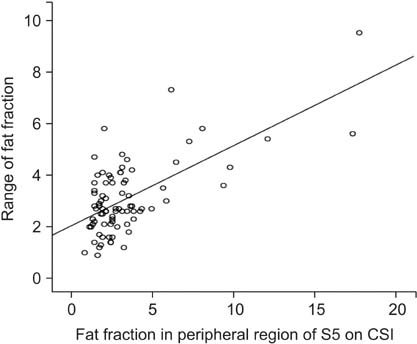
Fig. 3 The range of the estimated fat fraction increased with the estimated fat fraction in the peripheral region of segment 5 (S5) on CSI (P < 0.001). CSI, magnetic resonance chemical shift imaging.
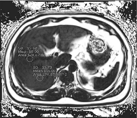
Fig. 4 Uneven distribution of fat in the liver. It measures 11.5% in segment 7 and 5% in segment 8. SD, standard deviation.
Reference
-
1. Wieckowska A, McCullough AJ, Feldstein AE. Noninvasive diagnosis and monitoring of nonalcoholic steatohepatitis: present and future. Hepatology. 2007; 46:582–589.2. Schwimmer JB, Deutsch R, Kahen T, Lavine JE, Stanley C, Behling C. Prevalence of fatty liver in children and adolescents. Pediatrics. 2006; 118:1388–1393.3. Adam R, Bismuth H, Diamond T, Ducot B, Morino M, Astarcioglu I, et al. Effect of extended cold ischaemia with UW solution on graft function after liver transplantation. Lancet. 1992; 340:1373–1376.4. Guiu B, Petit JM, Loffroy R, Ben Salem D, Aho S, Masson D, et al. Quantification of liver fat content: comparison of triple-echo chemical shift gradient-echo imaging and in vivo proton MR spectroscopy. Radiology. 2009; 250:95–102.5. Cho JY, Suh KS, Kwon CH, Yi NJ, Cho SY, Jang JJ, et al. The hepatic regeneration power of mild steatotic grafts is not impaired in living-donor liver transplantation. Liver Transpl. 2005; 11:210–217.6. Ratziu V, Charlotte F, Heurtier A, Gombert S, Giral P, Bruckert E, et al. Sampling variability of liver biopsy in nonalcoholic fatty liver disease. Gastroenterology. 2005; 128:1898–1906.7. Lee SW, Park SH, Kim KW, Choi EK, Shin YM, Kim PN, et al. Unenhanced CT for assessment of macrovesicular hepatic steatosis in living liver donors: comparison of visual grading with liver attenuation index. Radiology. 2007; 244:479–485.8. Park SH, Kim PN, Kim KW, Lee SW, Yoon SE, Park SW, et al. Macrovesicular hepatic steatosis in living liver donors: use of CT for quantitative and qualitative assessment. Radiology. 2006; 239:105–112.9. Ma X, Holalkere NS, Kambadakone RA, Mino-Kenudson M, Hahn PF, Sahani DV. Imaging-based quantification of hepatic fat: methods and clinical applications. Radiographics. 2009; 29:1253–1277.10. Henninger B, Kremser C, Rauch S, Eder R, Judmaier W, Zoller H, et al. Evaluation of liver fat in the presence of iron with MRI using T2* correction: a clinical approach. Eur Radiol. 2013; 23:1643–1649.11. Peng XG, Ju S, Qin Y, Fang F, Cui X, Liu G, et al. Quantification of liver fat in mice: comparing dual-echo Dixon imaging, chemical shift imaging, and 1H-MR spectroscopy. J Lipid Res. 2011; 52:1847–1855.12. Hatta T, Fujinaga Y, Kadoya M, Ueda H, Murayama H, Kurozumi M, et al. Accurate and simple method for quantification of hepatic fat content using magnetic resonance imaging: a prospective study in biopsy-proven nonalcoholic fatty liver disease. J Gastroenterol. 2010; 45:1263–1271.13. Joe E, Lee JM, Kim KW, Lee KB, Kim SJ, Baek JH, et al. Quantification of hepatic macrosteatosis in living, related liver donors using T1-independent, T2*-corrected chemical shift MRI. J Magn Reson Imaging. 2012; 36:1124–1130.14. Hamer OW, Aguirre DA, Casola G, Lavine JE, Woenckhaus M, Sirlin CB. Fatty liver: imaging patterns and pitfalls. Radiographics. 2006; 26:1637–1653.15. Ishizaka K, Oyama N, Mito S, Sugimori H, Nakanishi M, Okuaki T, et al. Comparison of 1H MR spectroscopy, 3-point DIXON, and multi-echo gradient echo for measuring hepatic fat fraction. Magn Reson Med Sci. 2011; 10:41–48.16. Kang BK, Yu ES, Lee SS, Lee Y, Kim N, Sirlin CB, et al. Hepatic fat quantification: a prospective comparison of magnetic resonance spectroscopy and analysis methods for chemical-shift gradient echo magnetic resonance imaging with histologic assessment as the reference standard. Invest Radiol. 2012; 47:368–375.17. Bashir MR, Merkle EM, Smith AD, Boll DT. Hepatic MR imaging for in vivo differentiation of steatosis, iron deposition and combined storage disorder: single-ratio in/opposed phase analysis vs. dual-ratio Dixon discrimination. Eur J Radiol. 2012; 81:e101–e109.18. Kim SH, Lee JM, Han JK, Lee JY, Lee KH, Han CJ, et al. Hepatic macrosteatosis: predicting appropriateness of liver donation by using MR imaging: correlation with histopathologic findings. Radiology. 2006; 240:116–129.19. Lee SS, Park SH, Kim HJ, Kim SY, Kim MY, Kim DY, et al. Non-invasive assessment of hepatic steatosis: prospective comparison of the accuracy of imaging examinations. J Hepatol. 2010; 52:579–585.20. Meisamy S, Hines CD, Hamilton G, Sirlin CB, McKenzie CA, Yu H, et al. Quantification of hepatic steatosis with T1independent, T2-corrected MR imaging with spectral modeling of fat: blinded comparison with MR spectroscopy. Radiology. 2011; 258:767–775.
- Full Text Links
-
- Actions
-
Cited
- CITED
-
- Close
- Share
-
- Similar articles
-
- Correlation Between Vertebral Marrow Fat Fraction Measured Using Dixon Quantitative Chemical Shift MRI and BMD Value on Dual-energy X-ray Absorptiometry
- Usefulness of preoperative magnetic resonance spectroscopy in improving the safety of a living liver donor
- Hepatic Artery Reconstruction Using the Right Gastroepiploic Artery for Hepatic Artery Inflow in a Living Donor Liver Transplantation
- Evaluation of Portal Venous Velocity with Doppler Ultrasound in Patients with Nonalcoholic Fatty Liver Disease
- Cutoff Values for Diagnosing Hepatic Steatosis Using Contemporary MRI-Proton Density Fat Fraction Measuring Methods
