Anat Cell Biol.
2013 Mar;46(1):79-81. 10.5115/acb.2013.46.1.79.
Unilateral duplication of vas deferens: a cadaveric case report
- Affiliations
-
- 1Department of Anatomy, Melaka Manipal Medical College, Manipal University, Manipal, Karnataka, India.
- 2Department of Anatomy, Kasturba Medical College, Manipal University, Manipal, Karnataka, India. kumar.mr@manipal.edu
- KMID: 2046760
- DOI: http://doi.org/10.5115/acb.2013.46.1.79
Abstract
- Duplication of vas deferens is a rare congenital anomaly. All previously reported cases of this rare anomaly were identified during procedures such as orchiepexy, inguinal hernia repair, vasectomy, varicocoelectomy, and radical prostatectomy. Here, we report a case of unilateral duplicated vas deferens noted in an adult cadaver during regular dissection for medical students. The right spermatic cord contained 2 separate and completely developed cord-like structures. Both cords communicated separately with the tail of the epididymis. When traced cranially, both traversed the inguinal canal as content of the spermatic cord and finally fused at the level of the deep inguinal ring. No other variations were found in the testis or epididymis, and no variations were seen in the left spermatic cord. In addition, no associated renal abnormalities were noted.
Keyword
MeSH Terms
Figure
-
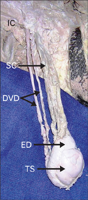
Fig. 1 Anterior view of the dissected spermatic cord showing its contents. Note 2 parallel vas deferens separated from the rest of the spermatic cord contents, running towards the inguinal canal. DVD, duplicated vas deferens; ED, epididymis; IC, inguinal canal; SC, spermatic cord contents; TS, testis.

Fig. 2 Closer view of the origin of the vas deferens. Note the duplicated vas deferens originating from the tail of the epididymis. BED, body of epididymis; DVD, duplicated vas deferens; HED, head of epididymis; TED, tail of epididymis.
Reference
-
1. Standring S, Borley NR, Collins P, Crossman AR, Gatzoulis MA, Healy JC, Johnson D, Mahadevan V, Newell RL, Wigley CB. Gray's anatomy: the anatomical basis of clinical practice. 2008. 40th ed. London, Churchill Livingstone: Elsevier;1268–1269.2. Karaman A, Karaman I, Yağiz B, Cavuşoğlu YH. Partial duplication of vas deferens: How important is it? J Indian Assoc Pediatr Surg. 2010. 15:135–136.3. Akay F, Atug F, Turkeri L. Partial duplication of the vas deferens at the level of inguinal canal. Int J Urol. 2005. 12:773–775.4. Chintamani , Khandelwal R, Tandon M, Kumar Y. Isolated unilateral duplication of vas deferens, a surgical enigma: a case report and review of the literature. Cases J. 2009. 2:167.5. Pick TP, Howden R. Gray's anatomy. 1901. Commemorative ed. New York: Bounty Books.6. Binderow SR, Shah KD, Dolgin SE. True duplication of the vas deferens. J Pediatr Surg. 1993. 28:269–270.7. Carr R. Apparent bilateral duplication of the vas deferens. Br J Urol. 1993. 71:354.8. Liang MK, Subramanian A, Weedin J, Griffith DP, Awad SS. True duplication of the vas deferens: a case report and review of literature. Int Urol Nephrol. 2012. 44:385–391.9. Vohra S, Morgentaler A. Congenital anomalies of the vas deferens, epididymis, and seminal vesicles. Urology. 1997. 49:313–321.10. Mysorekar VR. Accessory vas deferens: a case report. Br J Urol. 1976. 48:82.
