Malignant Arrhythmia with Benign Tumour: Fibrolipoma of the Left Ventricle
- Affiliations
-
- 1Department of Cardiology, Government Medical College, Kozhikode, Kerala, India. drsajeerkt@gmail.com
- 2Department of Radiodiagnosis, Government Medical College, Kozhikode, Kerala, India.
- 3Department of Cardiothoracic Surgery, Government Medical College, Kozhikode, Kerala, India.
- KMID: 2045433
- DOI: http://doi.org/10.4250/jcu.2014.22.3.151
Abstract
- We report a case of young male referred for evaluation of recent onset recurrent syncope. Inhospital electrocardiogram revealed an episode of ventricular flutter which reverted spontaneously to sinus rhythm. Transthoracic echocardiogram showed hyperechoic mass in the left ventricle. For further tissue characterization a cardiac magnetic resonance imaging was done which revealed a left ventricular mass with predominant fat content. The tumor was surgically resected. Histopathological examination confirmed the diagnosis of cardiac fibrolipoma. The patient recovered and is currently asymptomatic.
MeSH Terms
Figure
-
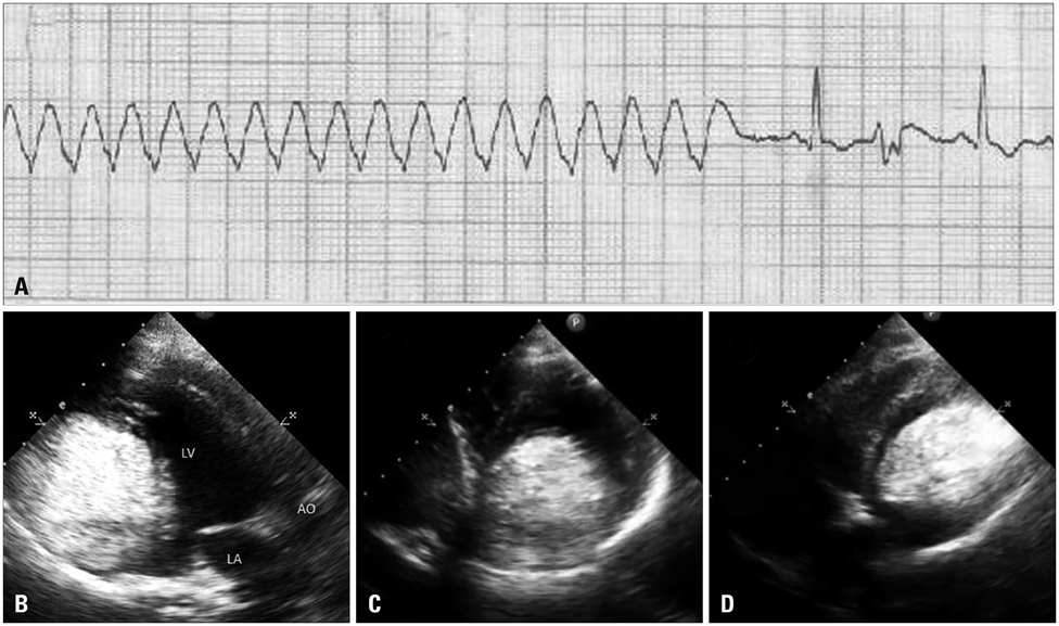
Fig. 1 A: Electrocardiogram rhythm strip showing ventricular flutter with spontaneous termination. Transthoracic echocardiogram showing hyper-echoic mass in left ventricular cavity arising form posterior wall (B: parasternal long axis view, C: parasternal short axis view, D: apical 4 chamber view). AO: aorta, LA: left atrium, LV: left ventricle.
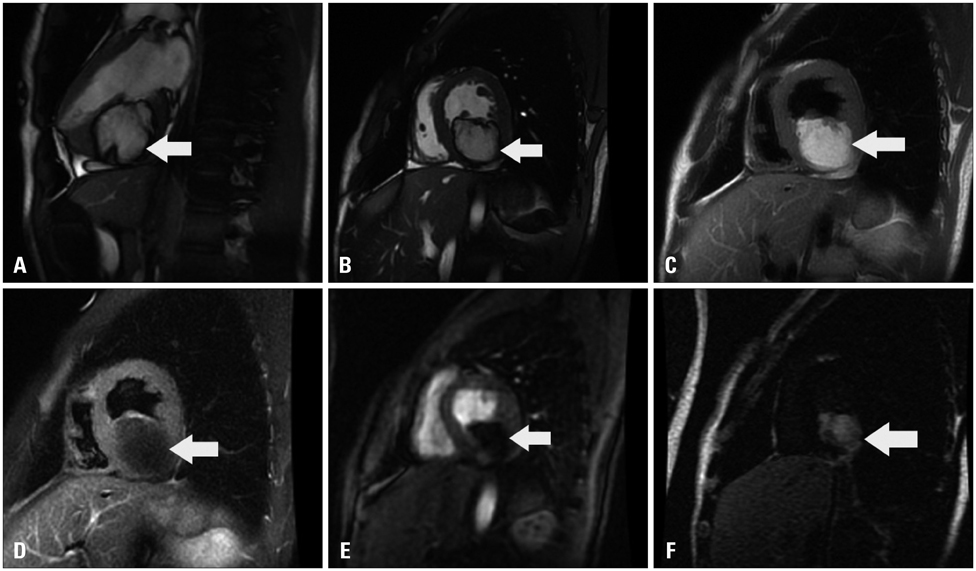
Fig. 2 Cardiac magnetic resonance imaging of left ventricular mass. A: T1-weighted image showing lobulated hyperintense lesion with peripheral hypointensity on saggital view. B: T1-weighted image-axial view. C: Double inversion recovery image showing hyperintense lesion indicating fat. D: Tripple inversion recovery image showing fat suppression. E: Complete suppression in signal intensity of fat on short tau inversion recovery sequence. F: Image showing post gadolinium delayed hypointense signal intensity in cardiac mass lesion suggestive of fibrous tissue.
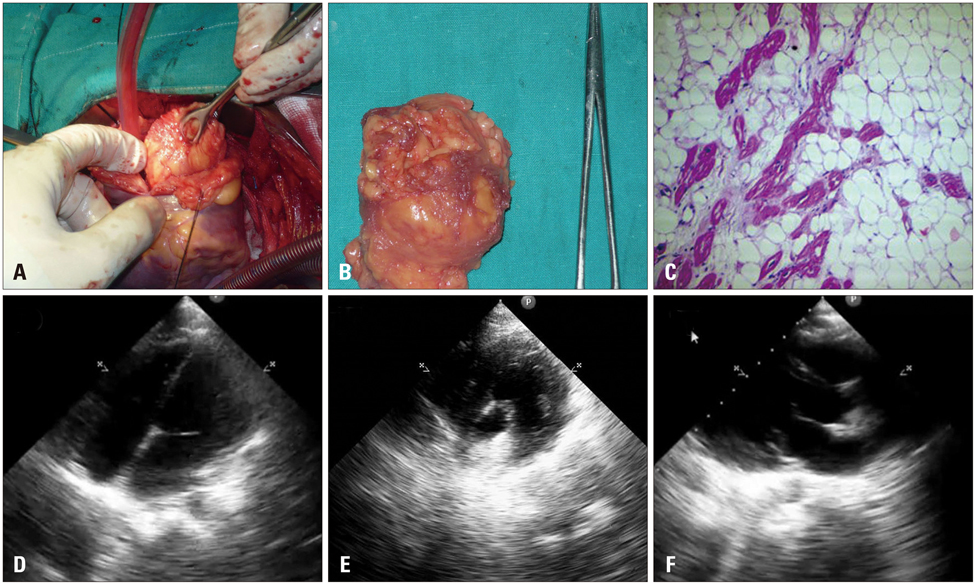
Fig. 3 A: Surgical picture showing the open left ventricle and the tumour being dissected. B: Gross specimen of the tumor. C: Histopathology showing mature fat cells with occasional fibrous connective tissue consistent with fibrolipoma (H&E stain, × 100 magnification). Post operative transthoracic echocardiogram showing complete clearance of left ventricular tumor (D: apical four chamber view, E: short axis view, F: parasternal long axis view).
Reference
-
1. Burke A, Virmani R. Tumours of the heart and great vessels. Atlas of tumor pathology, 3rd series, fascicle 16. Washington: Armed Forces Institute of Pathology;1996.2. Salanitri JC, Pereles FS. Cardiac lipoma and lipomatous hypertrophy of the interatrial septum: cardiac magnetic resonance imaging findings. J Comput Assist Tomogr. 2004; 28:852–856.3. Barberger-Gateau P, Paquet M, Desaulniers D, Chenard J. Fibrolipoma of the mitral valve in a child. Clinical and echocardiographic features. Circulation. 1978; 58:955–958.
Article4. Stratemann S, Dzurik Y, Fish F, Parra D. Left ventricular cardiac fibroma in a child presenting with ventricular tachycardia. Pediatr Cardiol. 2008; 29:223–226.
Article5. Ottaviani G, Rossi L, Ramos SG, Matturri L. Pathology of the heart and conduction system in a case of sudden death due to a cardiac fibroma in a 6-month-old child. Cardiovasc Pathol. 1999; 8:109–112.
Article6. Araoz PA, Mulvagh SL, Tazelaar HD, Julsrud PR, Breen JF. CT and MR imaging of benign primary cardiac neoplasms with echocardiographic correlation. Radiographics. 2000; 20:1303–1319.
Article7. Akram K, Hill C, Neelagaru N, Parker M. A left ventricular lipoma presenting as heart failure in a septuagenarian: a first case report. Int J Cardiol. 2007; 114:386–387.
Article
