J Korean Soc Radiol.
2015 Sep;73(3):147-157. 10.3348/jksr.2015.73.3.147.
Quantitative CT Assessment in Chronic Obstructive Pulmonary Disease Patients: Comparison of the Patients with and without Consistent Clinical Symptoms and Pulmonary Function Results
- Affiliations
-
- 1Department of Radiology, Soonchunhyang University Hospital, Seoul, Korea. jhhwang@schmc.ac.kr
- 2Bangbae GF Allergy Clinic, Seoul, Korea.
- 3Department of Radiology, Soonchunhyang University Bucheon Hospital, Bucheon, Korea.
- 4Department of Radiology, Soonchunhyang University Cheonan Hospital, Cheonan, Korea.
- 5Department of Preventive Medicine, Soonchunhyang University College of Medicine, Cheonan, Korea.
- KMID: 2043604
- DOI: http://doi.org/10.3348/jksr.2015.73.3.147
Abstract
- PURPOSE
We compared the clinical and quantitative CT measurement parameters between chronic obstructive pulmonary disease (COPD) patients with and without consistent clinical symptoms and pulmonary function results.
MATERIALS AND METHODS
This study included 60 patients having a clinical diagnosis of COPD, who underwent chest CT scan and pulmonary function tests. These 60 patients were classified into typical and atypical groups, which were further sub-classified into 4 groups, based on their dyspnea score and the result of pulmonary function tests [typical 1: mild dyspnea and pulmonary function impairment (PFI); typical 2: severe dyspnea and PFI; atypical 1: mild dyspnea and severe PFI; atypical 2: severe dyspnea and mild PFI]. Quantitative measurements of the CT data for emphysema, bronchial wall thickness and air-trapping were performed using software analysis. Comparative statistical analysis was performed between the groups.
RESULTS
The CT emphysema index correlated well with the results of the pulmonary functional test (typical 1 vs. atypical 1, p = 0.032), and the bronchial wall area ratio correlated with the dyspnea score (typical 1 vs. atypical 2, p = 0.033). CT air-trapping index also correlated with the results of the pulmonary function test (typical 1 vs. atypical 1, p = 0.012) and dyspnea score (typical 1 vs. atypical 2, p = 0.000), and was found to be the most significant parameter between the typical and atypical groups.
CONCLUSION
Quantitative CT measurements for emphysema and airways correlated well with the dyspnea score and pulmonary function results in patients with COPD. Air-trapping was the most significant parameter between the typical vs. atypical group of COPD patients.
MeSH Terms
Figure
-

Fig. 1 Findings of emphysema and color maps. Axial CT images (A, C) show diffuse centriacinar emphysema with low attenuation areas in both lungs. Color map images (B, D) show blue colored areas corresponding to emphysema lung (below -950 HU) and yellow colored areas corresponding to normal lung (above -900 HU). HU = Hounsfield units
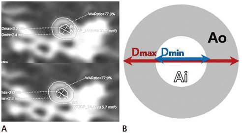
Fig. 2 Measurement of airway and schematic diagram. A. Short-axis image of the bronchus obtained from the curved multi-planar reformation is precisely perpendicular to the long-axis of the airway B. The schematic diagram of short-axis image of the bronchus shows the airway wall area (Ao), airway luminal area (Ai), outer diameter (Dmax), and inner diameter (Dmin).
Reference
-
1. Nishimura K, Izumi T, Tsukino M, Oga T. Dyspnea is a better predictor of 5-year survival than airway obstruction in patients with COPD. Chest. 2002; 121:1434–1440.2. Standards for the diagnosis and care of patients with chronic obstructive pulmonary disease. American Thoracic Society. Am J Respir Crit Care Med. 1995; 152(5 Pt 2):S77–S121.3. Vestbo J, Hurd SS, Agustí AG, Jones PW, Vogelmeier C, Anzueto A, et al. Global strategy for the diagnosis, management, and prevention of chronic obstructive pulmonary disease: GOLD executive summary. Am J Respir Crit Care Med. 2013; 187:347–365.4. Park KJ, Bergin CJ, Clausen JL. Quantitation of emphysema with three-dimensional CT densitometry: comparison with two-dimensional analysis, visual emphysema scores, and pulmonary function test results. Radiology. 1999; 211:541–547.5. Zaporozhan J, Ley S, Eberhardt R, Weinheimer O, Iliyushenko S, Herth F, et al. Paired inspiratory/expiratory volumetric thin-slice CT scan for emphysema analysis: comparison of different quantitative evaluations and pulmonary function test. Chest. 2005; 128:3212–3220.6. Mohamed Hoesein FA, de Jong PA, Lammers JW, Mali WP, Mets OM, Schmidt M, et al. Contribution of CT quantified emphysema, air trapping and airway wall thickness on pulmonary function in male smokers with and without COPD. COPD. 2014; 11:503–509.7. Hasegawa M, Nasuhara Y, Onodera Y, Makita H, Nagai K, Fuke S, et al. Airflow limitation and airway dimensions in chronic obstructive pulmonary disease. Am J Respir Crit Care Med. 2006; 173:1309–1315.8. Matsuoka S, Yamashiro T, Washko GR, Kurihara Y, Nakajima Y, Hatabu H. Quantitative CT assessment of chronic obstructive pulmonary disease. Radiographics. 2010; 30:55–66.9. Gibson PG, Simpson JL. The overlap syndrome of asthma and COPD: what are its features and how important is it? Thorax. 2009; 64:728–735.10. Lee YK, Oh YM, Lee JH, Kim EK, Lee JH, Kim N, et al. Quantitative assessment of emphysema, air trapping, and airway thickening on computed tomography. Lung. 2008; 186:157–165.11. Park HJ, Hwang JH. Quantification of emphysema with a three-dimensional chest CT scan: correlation with the visual emphysema scoring on chest CT, pulmonary function tests and dyspnea severity. J Korean Soc Radiol. 2011; 65:247–256.12. Tanaka N, Matsumoto T, Miura G, Emoto T, Matsunaga N, Ueda K, et al. Air trapping at CT: high prevalence in asymptomatic subjects with normal pulmonary function. Radiology. 2003; 227:776–785.13. Busacker A, Newell JD Jr, Keefe T, Hoffman EA, Granroth JC, Castro M, et al. A multivariate analysis of risk factors for the air-trapping asthmatic phenotype as measured by quantitative CT analysis. Chest. 2009; 135:48–56.14. Mets OM, van Hulst RA, Jacobs C, van Ginneken B, de Jong PA. Normal range of emphysema and air trapping on CT in young men. AJR Am J Roentgenol. 2012; 199:336–340.15. Newman KB, Lynch DA, Newman LS, Ellegood D, Newell JD Jr. Quantitative computed tomography detects air trapping due to asthma. Chest. 1994; 106:105–109.16. Van Tho N, Wada H, Ogawa E, Nakano Y. Recent findings in chronic obstructive pulmonary disease by using quantitative computed tomography. Respir Investig. 2012; 50:78–87.17. Madani A, Zanen J, de Maertelaer V, Gevenois PA. Pulmonary emphysema: objective quantification at multi-detector row CT--comparison with macroscopic and microscopic morphometry. Radiology. 2006; 238:1036–1043.18. Bommart S, Marin G, Bourdin A, Molinari N, Klein F, Hayot M, et al. Relationship between CT air trapping criteria and lung function in small airway impairment quantification. BMC Pulm Med. 2014; 14:29.19. Hogg JC, Chu F, Utokaparch S, Woods R, Elliott WM, Buzatu L, et al. The nature of small-airway obstruction in chronic obstructive pulmonary disease. N Engl J Med. 2004; 350:2645–2653.20. Nakano Y, Muro S, Sakai H, Hirai T, Chin K, Tsukino M, et al. Computed tomographic measurements of airway dimensions and emphysema in smokers. Correlation with lung function. Am J Respir Crit Care Med. 2000; 162(3 Pt 1):1102–1108.21. Lucidarme O, Coche E, Cluzel P, Mourey-Gerosa I, Howarth N, Grenier P. Expiratory CT scans for chronic airway disease: correlation with pulmonary function test results. AJR Am J Roentgenol. 1998; 170:301–307.22. Eda S, Kubo K, Fujimoto K, Matsuzawa Y, Sekiguchi M, Sakai F. The relations between expiratory chest CT using helical CT and pulmonary function tests in emphysema. Am J Respir Crit Care Med. 1997; 155:1290–1294.23. Kauczor HU, Hast J, Heussel CP, Schlegel J, Mildenberger P, Thelen M. CT attenuation of paired HRCT scans obtained at full inspiratory/expiratory position: comparison with pulmonary function tests. Eur Radiol. 2002; 12:2757–2763.24. Akira M, Toyokawa K, Inoue Y, Arai T. Quantitative CT in chronic obstructive pulmonary disease: inspiratory and expiratory assessment. AJR Am J Roentgenol. 2009; 192:267–272.25. Galbán CJ, Han MK, Boes JL, Chughtai KA, Meyer CR, Johnson TD, et al. Computed tomography-based biomarker provides unique signature for diagnosis of COPD phenotypes and disease progression. Nat Med. 2012; 18:1711–1715.26. Schroeder JD, McKenzie AS, Zach JA, Wilson CG, Curran-Everett D, Stinson DS, et al. Relationships between airflow obstruction and quantitative CT measurements of emphysema, air trapping, and airways in subjects with and without chronic obstructive pulmonary disease. AJR Am J Roentgenol. 2013; 201:W460–W470.
- Full Text Links
-
- Actions
-
Cited
- CITED
-
- Close
- Share
-
- Similar articles
-
- Clinical use of chest CT in chronic obstructive pulmonary diseases
- Quantitative CT Imaging in Chronic Obstructive Pulmonary Disease: Review of Current Status and Future Challenges
- Nutritional Assessment in Patients with Chronic Obstructive Pulmonary Disease
- Cor Pulmonale with Particular Reference to Chronic Obstructive Pulmonary Disease and Pulmonary Tuberculosis
- The Study on the Effects of a Respiratory Rehabilitation Program for COPD Patients
