Hemorrhagic Focal Nodular Hyperplasia in Young Men: A Case Report
- Affiliations
-
- 1Department of Diagnostic Radiology, College of Medicine, Dong-A University, Busan, Korea. risual@dau.ac.kr
- 2Department of Surgery, College of Medicine, Dong-A University, Busan, Korea.
- 3Department of Pathology, College of Medicine, Dong-A University, Busan, Korea.
- KMID: 2041940
- DOI: http://doi.org/10.3348/jksr.2014.70.4.291
Abstract
- In general, focal nodular hyperplasia is a hepatic lesion that most frequently affects the healthy women of reproductive age. Focal nodular hyperplasia lesions have a benign natural course; the majority of the cases remain asymptomatic and complications are rare. Spontaneous hemorrhage of focal nodular hyperplasia is a rare disease, and the hemorrhage in young men is even more uncommon. We report a rare case of spontaneous hemorrhage of focal nodular hyperplasia in a 19-year-old man.
MeSH Terms
Figure
-
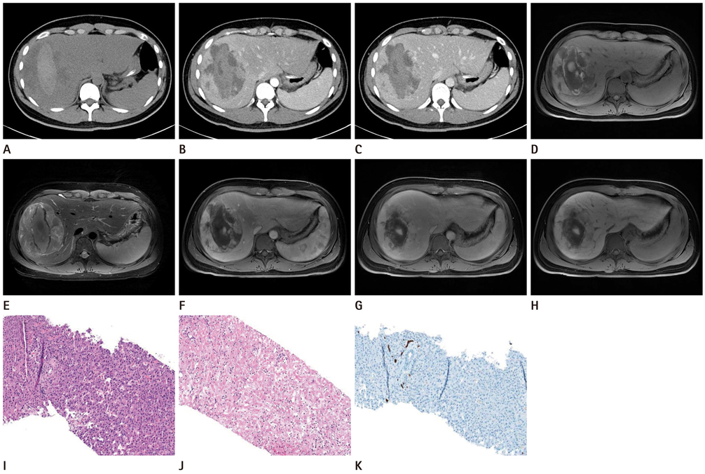
Fig. 1 19-year-old man presenting abdominal pain in upper abdomen. A. Axial CT image shows a large hepatic mass (about 12 × 8 cm in size) and central low density portion within the mass, considered as central necrosis and high density portion within the mass, considered as hemorrhagic portion. B, C. Axial images of contrast-enhanced CT show mild enhancement of peripheral portion of mass in arterial phase (B) and iso attenuation in portal phase (C). D. After 6 days, follow-up MR imaging was obtained. T1-weighted image shows heterogeneous mass (about 12 × 8 cm in size) in the right lobe and central high signal intensity and low signal intensity portion. The peripheral portion of the mass shows a isointense signal. E. T2-weighted image shows a hyperintense signal in the peripheral portion. F-H. Contrast-enhanced dynamic scan shows early mild enhancement in arterial phase (F) and a isointense signal in the portal (G), delayed, hepatobiliary phase (H). I, J. After liver needle biopsy, the specimen was obtained. Expansile proliferation of hepatocyte (I) and central hemorrhage (J) are shown in hematoxylin and eosin stain (× 100). K. Immunohistochemical staining of cytokeratin 7 (CK7). The result shows positive immunohistochemical staining of CK7 representing focal nodular hyperplasia.
Reference
-
1. Casillas VJ, Amendola MA, Gascue A, Pinnar N, Levi JU, Perez JM. Imaging of nontraumatic hemorrhagic hepatic lesions. Radiographics. 2000; 20:367–378.2. Ishak KG, Rabin L. Benign tumors of the liver. Med Clin North Am. 1975; 59:995–1013.3. Wanless IR, Mawdsley C, Adams R. On the pathogenesis of focal nodular hyperplasia of the liver. Hepatology. 1985; 5:1194–1200.4. Li T, Qin LX, Ji Y, Sun HC, Ye QH, Wang L, et al. Atypical hepatic focal nodular hyperplasia presenting as acute abdomen and misdiagnosed as hepatocellular carcinoma. Hepatol Res. 2007; 37:1100–1105.5. Becker YT, Raiford DS, Webb L, Wright JK, Chapman WC, Pinson CW. Rupture and hemorrhage of hepatic focal nodular hyperplasia. Am Surg. 1995; 61:210–214.6. Knowles DM 2nd, Casarella WJ, Johnson PM, Wolff M. The clinical, radiologic, and pathologic characterization of benign hepatic neoplasms Alleged association with oral contraceptives. Medicine (Baltimore). 1978; 57:223–237.7. Whelan TJ Jr, Baugh JH, Chandor S. Focal nodular hyperplasia of the liver. Ann Surg. 1973; 177:150–158.8. Goodwin MD, Dobson JE, Sirlin CB, Lim BG, Stella DL. Diagnostic challenges and pitfalls in MR imaging with hepatocyte-specific contrast agents. Radiographics. 2011; 31:1547–1568.9. Scalori A, Tavani A, Gallus S, La Vecchia C, Colombo M. Risk factors for focal nodular hyperplasia of the liver: an Italian case-control study. Am J Gastroenterol. 2002; 97:2371–2373.10. Mathieu D, Kobeiter H, Maison P, Rahmouni A, Cherqui D, Zafrani ES, et al. Oral contraceptive use and focal nodular hyperplasia of the liver. Gastroenterology. 2000; 118:560–564.
