Solitary Fibrous Tumor of the Ischiorectal Fossa : CT and MRI Findings
- Affiliations
-
- 1Department of Radiology, College of Medicine, Korea University, Korea. sahcha@kumc.or.kr
- KMID: 2041230
- DOI: http://doi.org/10.13104/jksmrm.2011.15.1.72
Abstract
- Solitary fibrous tumor (SFT) is a rare neoplasm, which is usually presented as a pleural based mass, but can also occur in unusual locations based on its mesenchymal origin. However, the radiologic features of SFT occurred in the ischiorectal fossa have been rarely reported. In this case, we describe the MRI findings in a case of a SFT involving the ischiorectal fossa of a 36-year-old man. The tumor appeared as homogeneous iso-signal intensity relative to the adjacent muscle on T1 weighted images, a mixed high signal intensity on the T2 weighted images, and heterogeneous enhancement following the administration of the contrast material.
Keyword
MeSH Terms
Figure
-
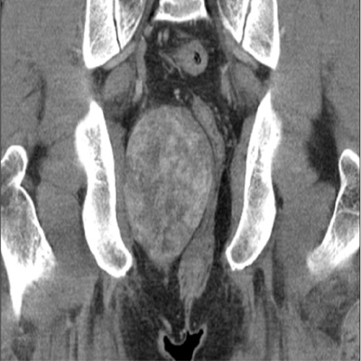
Fig. 1 Contrast enhanced coronal reformatted CT shows large, well defined mass with heterogeneous attenuation in the right ischiorectal fossa.
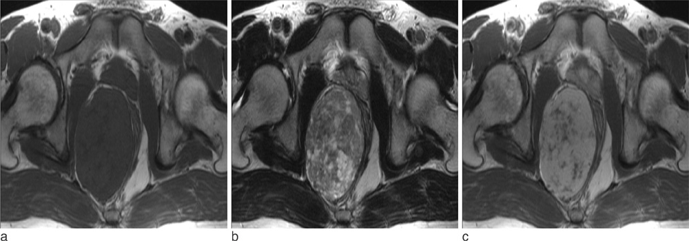
Fig. 2 Axial T1 weighted (a), T2 weighted (b), and contrast enhanced T1 weighted (c) images show soft tissue mass in right ischiorectal fossa. Mass exhibits homogeneous high signal intensity in (a) and heterogeneous high signal in (b), and heterogeneous enhancement in (c). The hyperintense area on T2-weighted images within mass revealed varying degree of enhancement.

Fig. 3 Sagittal contrasted enhanced T1 weighted images demonstrate multiple tubular structures arising from the subcutaneous fat of perineum. The low signal intensity of these structures, caused by a flow void effect, is suggestive of collateral feeding vessels (arrow).
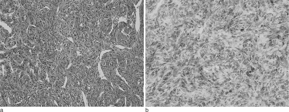
Fig. 4 (a) The tumor is composed of short spindle cells with abundant stromal collagen bundles and ectactic vessels (hematoxylin and eosin staining, ×200). (b) The tumor cells are diffuse strong positive for CD34 staining (hematoxylin and eosin staining, ×200).
Reference
-
1. Gengler C, Guillou L. Solitary fibrous tumour and haemangiopericytoma: evolution of a concept. Histopathology. 2006. 48:63–74.2. Goodlad JR, Fletcher CD. Solitary fibrous tumour arising at unusual sites: analysis of a series. Histopathology. 1991. 19:515–522.3. Chun HJ, Byun JY, Jung SE, Kim KH, Shinn KS. Benign solitary fibrous tumour of the pre-sacral space: MRI findings. Br J Radiol. 1998. 71:677–679.4. Wignall OJ, Moskovic EC, Thway K, Thomas JM. Solitary fibrous tumors of the soft tissues: review of the imaging and clinical features with histopathologic correlation. AJR Am J Roentgenol. 2010. 195:W55–W62.5. Rosenkrantz AB, Hindman N, Melamed J. Imaging appearance of solitary fibrous tumor of the abdominopelvic cavity. J Comput Assist Tomogr. 2010. 34:201–205.6. Vossough A, Torigian DA, Zhang PJ, Siegelman ES, Banner MP. Extrathoracic solitary fibrous tumor of the pelvic peritoneum with central malignant degeneration on CT and MRI. J Magn Reson Imaging. 2005. 22:684–686.7. Gold JS, Antonescu CR, Hajdu C, Ferrone CR, Hussain M, Lewis JJ, et al. Clinicopathologic correlates of solitary fibrous tumors. Cancer. 2002. 94:1057–1068.8. Vallat-Decouvelaere AV, Dry SM, Fletcher CD. Atypical and malignant solitary fibrous tumors in extrathoracic locations: Evidence of their comparability to intra-thoracic tumors. Am J Surg Pathol. 1998. 22:1501–1511.9. Jeong AK, Lee HK, Kim SY, Cho KJ. Solitary fibrous tumor of the parapharyngeal space: MR imaging findings. AJNR Am J Neuroradiol. 2002. 23:473–475.10. Kim HJ, Lee HK, Seo JJ, Shin JH, Jeong AK, Lee JH, et al. MR imaging of solitary fibrous tumors in the head and neck. Korean J Radiol. 2005. 6:136–142.11. Kakihara D, Yoshimitsu K, Eto M, Matsuura S, Honda H. MRI of retroperitoneal solitary fibrous tumor in the suprarenal region. AJR Am J Roentgenol. 2007. 188:W512–W514.12. Casas JD, Balliu E, Sánchez MC, Mariscal A. Benign solitary fibrous tumor of the retroperitoneum: radiological features. CMIG Extra: Cases. 2004. 28:50–53.13. Hide IG, Baudouin CJ, Murray SA, Malcolm AJ. Giant ancient schwannoma of the pelvis. Skeletal Radiol. 2000. 29:538–542.14. Totterman S, Reitamo JJ. Desmoid tumour: an angiographic study of five cases. Br J Radiol. 1979. 52:936–941.15. Llauger J, Palmer J, Perez C, Monill J, Ribe J, Moreno A. The normal and pathologic ischiorectal fossa at CT and MR imaging. Radiographics. 1998. 18:61–82. quiz 146.16. Lee JC, Thomas JM, Phillips S, Fisher C, Moskovic E. Aggressive fibromatosis: MRI features with pathologic correlation. AJR Am J Roentgenol. 2006. 186:247–254.17. Rha SE, Byun JY, Jung SE, Chun HJ, Lee HG, Lee JM. Neurogenic tumors in the abdomen: tumor types and imaging characteristics. Radiographics. 2003. 23:29–43.
- Full Text Links
-
- Actions
-
Cited
- CITED
-
- Close
- Share
-
- Similar articles
-
- Imaging Findings of a Solitary Fibrous Tumor in Pancreas: A Case Report
- Solitary Fibrous Tumor of the Adrenal Gland: A Case Report
- Malignant Solitary Fibrous Tumor in the Perianal Region
- An Ancillary CT Finding of Intrapulmonary Solitary Fibrous Tumor: A Case Report
- Aggressive AngiOmYxoma Occuring in Ischiorectal Fossa: A case report
